
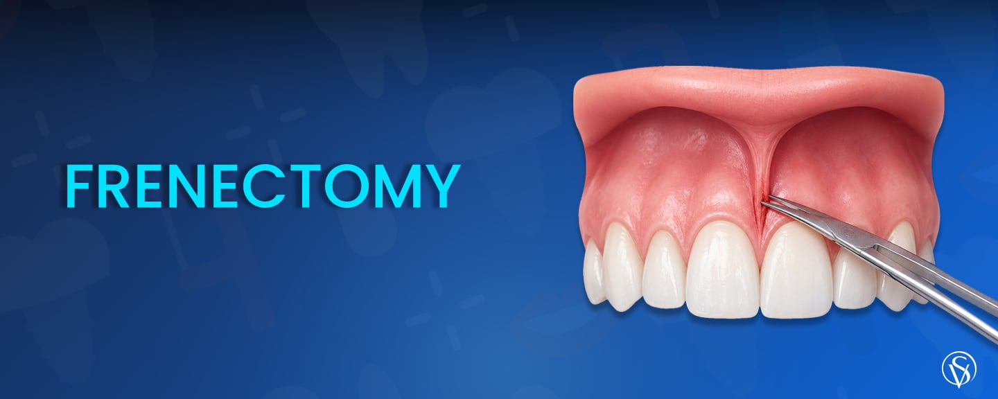
A frenectomy is a minor oral surgical procedure that releases or removes a frenulum, the small fold of soft tissue anchoring the lip or the tongue to the gum or the floor of the mouth. Two types account for almost all cases. A labial frenectomy treats the fold connecting the lip to the gum, and a lingual frenectomy releases a restrictive tongue attachment known as tongue-tie. The procedure is indicated when frenal tension causes a midline gap, gum recession, plaque retention, denture instability, or restricted lip and tongue movement. It is performed in a single appointment under local anesthesia with either a scalpel or a diode laser, and the surgical part takes only a few minutes. In children, timing matters more than technique, because operating before the permanent canines erupt raises the risk of regrowth and orthodontic relapse. The benefits are functional rather than cosmetic, and soft tissue healing runs across the first two to four weeks.
Key Points
- A frenectomy releases or removes a frenulum. The two types are labial, treating the lip attachment, and lingual, releasing a tongue-tie.
- It is a single appointment under local anesthesia, and the surgical part takes only a few minutes.
- A diode laser lowers early pain, bleeding, and operating time, 341 against 675 seconds, but the final result matches the scalpel.
- In children, surgery is deferred until the permanent canines erupt, ages 9 to 12, to avoid regrowth and orthodontic relapse.
- The benefits are functional, not cosmetic. A frenectomy does not close a gap or straighten teeth on its own.
- Most discomfort falls in the first week, and full soft tissue healing takes 3 to 4 weeks.
What Is a Frenectomy?
A frenectomy is the surgical removal or release of a frenulum together with its attachment to the underlying tissue. It is performed by a dentist, a periodontist, or an oral surgeon and is classed as minor oral surgery rather than a major operation. The aim in most cases is functional rather than cosmetic, since the frenulum is removed to relieve the tension it places on the gum, the lip, or the tongue.
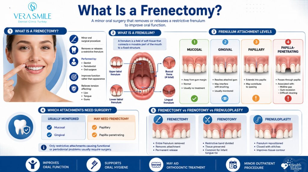
A frenulum is a fold of mucous membrane and connective tissue, sometimes containing muscle fibres, that tethers a mobile structure to a fixed one. The oral cavity contains seven frena, comprising the maxillary labial frenum, four buccal frena, the lingual frenum, and the mandibular labial frenum (Sobouti et al., 2024). Not every frenulum causes a problem. What matters is how far down it inserts, and four attachment levels are recognised.
The 4 Frenelum Attachment Levels
- Mucosal: Inserts into the mucosa well away from the gum margin and causes no clinical problem.
- Gingival: Reaches the attached gum tissue and may begin to interfere with brushing.
- Papillary: Extends into the interdental papilla between the two front teeth.
- Papilla-penetrating: Thick fibrous tissue passes through the papilla to the palatal side, and this level is the one most strongly linked to a midline gap, gum recession, and poor hygiene access.
Only the papilla-related attachments justify surgery. A mucosal or gingival attachment is monitored rather than operated on, and in growing children the attachment often shifts on its own, because the maxillary labial frenum migrates toward the mucogingival junction as the upper jaw develops (Marr et al., 2025). Three related terms are often confused, and they describe three different amounts of surgery.
| Frenectomy | Frenotomy | Frenuloplasty |
|---|---|---|
| Complete excision of the frenulum with its attachment to the periosteum | A simple release incision that divides the band without removing tissue | Repositions the frenulum and closes the wound with sutures |
| Used for persistent labial and lingual restrictions | Most often used for infant tongue-tie | Allows control of vestibule depth and final tissue contour |
What Are the Types of Frenectomy?
Frenectomy is classified by which frenulum is treated, and two types account for almost all cases performed. The distinction matters because each involves different anatomy, a different age group, and a different reason for treatment. The two types are:
- Labial frenectomy: Removal or release of the fold joining the upper or lower lip to the gum, most commonly the maxillary labial frenum behind the two upper front teeth, and most often carried out in adolescents and adults.
- Lingual frenectomy: Release of a short or tight lingual frenulum that restricts tongue movement, performed to address feeding difficulty in infants or restricted tongue mobility later in life.
Both procedures share the same surgical principle, which is relief of tension at the point of attachment. Where they differ is in who raises the concern. A labial case is most often identified by a dentist or an orthodontist during treatment planning, whereas a lingual case is frequently raised first by a parent or a lactation consultant. Infant and childhood cases are managed within pediatric dentistry, where feeding, speech development, and jaw growth are assessed together rather than in isolation.
Reported prevalence of tongue-tie varies widely, and the reason is diagnostic rather than biological. A meta-analysis of 24,536 infants under one year of age found a pooled ankyloglossia prevalence of 8% (95% CI 6% to 10%), with 7% in males and 4% in females (Hill et al., 2021). A larger meta-analysis across 71 studies reported an overall prevalence of 5% (95% CI 4% to 5%), rising to 7% in infants, with figures ranging from 2% to 20% depending purely on which assessment tool the examiners used (Cruz et al., 2022). The same baby can be classified as having ankyloglossia under one protocol and not under another, so a diagnosis is only as reliable as the assessment tool behind it.
Why Would You Need a Frenectomy?
A frenectomy is needed when an abnormally positioned frenulum causes a functional or periodontal problem that will not resolve on its own. The presence of a visible frenulum is not in itself an indication for surgery, and most frena never need treatment. What triggers the decision is evidence of tension being transmitted to the gum, the teeth, or a prosthesis. Six indications account for the majority of cases.
- Midline diastema: A papilla-penetrating frenulum holds the two upper front teeth apart and can reopen the gap after orthodontic closure.
- Gum recession: Repeated muscle pull on the gingival margin displaces the tissue apically and exposes the root surface.
- Plaque retention and food impaction: A thick attachment traps debris and blocks brush access, producing localised gum inflammation.
- Denture instability: A prominent frenulum interferes with the seating and retention of dentures in adults.
- Restricted lip or tongue movement: Limited mobility affects feeding in infants and oral clearance at any age.
- Orthodontic relapse: Residual frenal tension after treatment pulls closed teeth back toward their original position.
These indications overlap more often than they appear to. A single papilla-penetrating attachment can produce a gap, recession, and hygiene difficulty at the same time, which is why assessment looks at the whole picture rather than one symptom. Where denture retention is the concern, the frenectomy is planned alongside the fit of the Dentures so the flange seats without being displaced by lip movement.
Clinic Note: Frenal tension is assessed at the consultation with a simple retraction test. The upper lip is lifted and pulled outward while the interdental papilla is observed. If the papilla blanches, moves, or the gum margin visibly lifts away from the tooth, the attachment is transmitting force to the periodontium and is recorded as clinically significant. If the tissue stays still, the frenulum is documented and monitored rather than treated.
Who Is Not a Candidate for a Frenectomy?
Several groups are not treated surgically, either because the problem is likely to resolve without intervention or because another condition must be addressed first. Declining or deferring surgery in these cases is a clinical decision, not a delay. Four categories apply.
- Children before permanent canine eruption: Surgery is deferred, because the frenulum migrates apically with jaw growth and early surgery carries a regrowth risk.
- Cases with unexplored airway or developmental factors: Airway obstruction and developmental delay are ruled out before a frenulum is accepted as the cause of a feeding or speech concern (Marr et al., 2025).
- Uncontrolled bleeding disorders or anticoagulant therapy: Surgery waits for haematological assessment and medical clearance.
- Active infection at the surgical site: Local infection is resolved before any incision is made.
None of these is a permanent exclusion. A child deferred at eight years old may be a straightforward candidate at twelve, and a patient on anticoagulants can proceed once medical clearance is in place. What each category has in common is that operating first would either be unnecessary or would make the outcome less predictable.
What Are the Benefits of a Frenectomy?
The benefits of a frenectomy follow directly from relieving frenal tension, and they are functional rather than cosmetic. What a patient gains depends on which problem prompted the surgery, so the outcome for an infant with a feeding difficulty looks nothing like the outcome for an adult with an unstable denture. The gains are modest and specific. Six benefits are:
- Reduced frenal pull on the gum margin: Removing the attachment stops the repeated traction that displaces gingival tissue.
- Easier oral hygiene: Brush access to the midline improves once the obstructing band is gone, which reduces plaque retention.
- Improved denture retention: A denture flange seats without being displaced by lip movement.
- Support for orthodontic stability: Excising fibres under tension after diastema closure reduces the pull driving relapse.
- Easier feeding in infants: Releasing a restrictive lingual frenulum can improve tongue elevation for latch.
- Increased lip or tongue mobility: Range of movement improves where it was mechanically limited.
Each of these is a mechanical result of removing tension, which is why they are reliable within their limits and unreliable outside them. A frenectomy will not close a gap by itself, straighten teeth, or regenerate gum tissue that has already receded. Where recession or a diastema is already established, the frenectomy removes the cause while a separate procedure addresses the damage.
Does a frenectomy improve speech?
No. A frenectomy on its own is not a speech treatment. It can increase the range of tongue movement where that movement was mechanically restricted, but the articulation habits formed while the tongue was restricted do not change because the restriction was released. Where speech is the concern, surgery is one component of a plan that includes speech and language therapy, and the therapy is what produces the articulation change (Marr et al., 2025).
What Are the Risks and Side Effects of a Frenectomy?
Frenectomy is a low-risk procedure, and most side effects are mild, expected, and short-lived. The surgical field is small, the tissue is thin, and the wound is shallow, which keeps the complication profile narrow. Even so, patients should know what a normal recovery feels like so they can recognise the point at which it stops being normal. Six effects are:
- Swelling and soreness: Both peak in the first 48 hours, and no swelling was recorded in either the laser or the scalpel group by day 7 (Yousefimanesh et al., 2024).
- Minor bleeding: Oozing on the day of surgery is expected and is more common after scalpel technique than after laser (Sobouti et al., 2024).
- Discomfort in chewing and speaking: Reported through the first 7 days and more pronounced after scalpel technique (Sobouti et al., 2024).
- Scar tissue: A small band of fibrous tissue can form at the healed site.
- Temporary numbness: Sensation at the site can be altered for a short period after the anesthetic wears off.
- Infection: Uncommon, given the small wound and the blood supply of oral mucosa.
What separates a normal recovery from a complication is the direction of travel. Discomfort that is improving day by day is expected, while discomfort that is worsening after day three is not.
Warning: Contact the clinic the same day if you develop a fever, swelling that spreads beyond the surgical site or into the face, bleeding that does not stop after 10 minutes of firm pressure, or pain that worsens after the third day rather than improving. These are not features of normal healing.
Can a frenulum grow back after a frenectomy?
No. A frenectomy performed completely and at the right time does not result in reattachment. A three-arm randomized clinical trial recorded no frenum reattachment in any group, whether or not a periosteal horizontal incision was included (Sezgin et al., 2020). Regrowth, when it occurs, is linked to timing rather than technique: surgery carried out before the permanent maxillary canines erupt is associated with regrowth, and incomplete excision that leaves fibres attached to the periosteum is the other mechanism (Marr et al., 2025).
How Is a Frenectomy Performed?
A frenectomy is performed in a single appointment under local anesthesia, and the surgical part takes only a few minutes. The frenulum is detached from its insertion, the tension is released, and the wound is either closed with sutures or left to heal openly depending on the technique used. Four stages make up the appointment.
- Examination and planning: The attachment level is recorded, the retraction test is repeated, and the technique is selected.
- Local anesthesia: The area is numbed with local infiltration, and the patient stays awake throughout.
- Release or excision: The frenulum is divided or removed along with its attachment to the periosteum, and adjacent tissue is repositioned where the technique calls for it.
- Closure and healing: Sutures are placed in scalpel cases, laser wounds are commonly left to granulate, and post-operative instructions are given before the patient leaves.
Local anesthesia covers the great majority of labial and lingual cases in adults, adolescents, and cooperative children, with no fasting or recovery period needed. General anesthesia or sedation is reserved for a narrow group, principally very young children who cannot cooperate with an awake procedure and may require Sedation dentistry. In newborns, a simple frenotomy for tongue-tie is frequently performed without any anesthesia, because the release is a single shallow incision through a thin avascular band.
What Is the Difference Between Laser and Conventional Frenectomy?
Laser and scalpel frenectomy achieve the same surgical result but differ in operating time, bleeding, early pain, and how the wound is managed. Two randomized controlled trials supply the figures: one enrolled 174 patients in three groups of 58, assessed at day 7 and day 30 (Sobouti et al., 2024), and the second enrolled 26 patients with papillary or papilla-penetrating attachments (Yousefimanesh et al., 2024). The diode laser used in these procedures is one instrument option offered within Laser dentistry. The comparison below draws on both trials.
| Aspect | Diode laser | Scalpel |
|---|---|---|
| Mean surgery duration | 341 seconds | 675 seconds |
| Intraoperative bleeding | Significantly lower | Higher, requires haemostasis |
| Pain at day 1 and day 7 | Significantly lower | Higher |
| Chewing and speaking discomfort | Significantly lower at day 7 | Higher in the first week |
| Swelling score at day 1 | 0.61 mean | 1.30 mean |
| Analgesic tablets taken | 5.53 mean | 9.76 mean |
| Tissue healing at day 7 | Better than scalpel | Slower at day 7 |
| Sutures | Commonly not required | Placed in most cases |
| Reattachment at follow-up | None recorded | None recorded |
The advantage the laser holds is concentrated in the first week rather than in the final result. Healing scores favoured the laser at 1 week, 3.92 against 2.61, but the difference had disappeared by 1 month (Yousefimanesh et al., 2024). Within the diode group, the 445 nm wavelength outperformed 980 nm on pain and healing (Sobouti et al., 2024), and a separate trial found no reattachment after either technique (Sezgin et al., 2020). Instrument choice is therefore a question of early comfort, operating time, and case complexity rather than long-term success.
When Should a Frenectomy Be Performed?
A frenectomy in a child should be deferred until the permanent maxillary canines have erupted, between the ages of 9 and 12, and where it forms part of orthodontic care it is carried out after the diastema has been closed. Operating earlier is associated with regrowth, surgical complications, and relapse, and there is insufficient evidence to support early surgical intervention (Marr et al., 2025). Sequencing the surgery after tooth alignment is completed in Orthodontics removes the residual tension at the point where it would otherwise pull the teeth apart again.
The reasoning is developmental. The maxillary labial frenum sits low on the alveolar crest in infancy and migrates apically as the maxilla grows, so a conservative approach of monitoring allows the anatomy to correct itself in a substantial proportion of cases (Marr et al., 2025). Removing tissue before that migration has finished means operating on an attachment that was going to move anyway, and the regrown tissue that follows is often thicker and less predictable than the original. Once the teeth are together, the fibres still under tension are visible and can be excised completely, whereas operating on an open gap leaves the surgeon guessing which fibres will matter later.
Warning: A visible frenulum in a young child is not on its own a reason for surgery. Frenectomy performed before the permanent canines erupt carries a documented risk of regrowth, complications, and orthodontic relapse (Marr et al., 2025). Any recommendation for early surgery should be accompanied by a specific functional finding, and airway and developmental causes should be excluded first.
What Is Recovery Like After a Frenectomy?
Recovery from a frenectomy is straightforward, with the first week accounting for nearly all of the discomfort and the following weeks accounting for the tissue remodelling. Most patients return to work or school the same day or the next. The healing checkpoints used in clinical trials are day 7 and day 30, and these are useful markers for patients as well (Sobouti et al., 2024). Recovery divides into three phases.
- First 24 hours: Mild oozing, numbness wearing off, and peak swelling, with cold compresses and a soft diet from this point.
- Days 2 to 7: Soreness and discomfort in chewing and speaking, both declining daily, with sutures removed toward the end of this period where placed.
- Weeks 2 to 4: Epithelialisation completes and the tissue contour settles, while any prescribed stretching or mobility exercises continue.
Adherence during the second and third phases determines the final result more than anything that happens in the operating chair. Wounds left to heal openly close by granulation, which looks worse before it looks better and alarms patients who were not warned. Surface healing takes 1 to 2 weeks and full soft tissue remodelling takes 3 to 4 weeks, which is why the site can look slightly raised or pale at two weeks and normal at four. A review appointment confirms complete healing and, in orthodontic cases, confirms that the closed diastema is holding.
Patient Tips: Apply a cold compress in 20 minute intervals during the first 24 hours to limit swelling.Brush the surrounding teeth normally but angle the brush away from the wound for the first week.Rinse gently with warm salt water from the second day rather than the first, since early vigorous rinsing disturbs the clot.Carry out any stretching or tongue-mobility exercises exactly as prescribed, because they prevent fibrous tightening as the tissue contracts.
What Can You Eat After a Frenectomy?
Diet is restricted for the first few days and returns to normal within a week. The aim is to avoid mechanical disturbance of the wound and chemical irritation of exposed tissue, both of which prolong soreness without causing lasting harm. Food choice matters most in the first 48 hours, when the clot is least stable.
- Cool soft foods for the first 48 hours: Yoghurt, smoothies, mashed vegetables, and soft eggs place no load on the wound.
- Lukewarm rather than hot: Heat increases blood flow to the site and can restart oozing on the first day.
- Avoid crunchy and sharp textures: Crisps, toast crusts, nuts, and seeds can catch the healing tissue directly.
- Avoid spicy and acidic foods: Chilli, citrus, and vinegar-based dressings sting exposed granulation tissue.
- Avoid straws and vigorous suction: Negative pressure disturbs a fresh wound in the first 24 hours.
Most patients move back to a normal diet between day 4 and day 7, guided by comfort rather than by a fixed rule. Chewing on the opposite side and taking smaller mouthfuls covers most of the remaining restriction.
FAQ
Yes. A papilla-penetrating frenulum can hold the central incisors apart and can pull them apart again after orthodontic treatment has closed the space, because the fibrous band passes between the roots and resists the teeth being brought together. Most midline gaps in children are a normal developmental stage that closes on its own as the permanent canines erupt, so a gap in a seven-year-old is rarely evidence of a frenulum problem (Marr et al., 2025).
Yes. A frenulum attaching at or above the gum margin transmits muscular pull to the gingiva each time the lip moves, and that repeated traction can displace the margin and expose root surface over time. Recession from frenal pull is localised and asymmetric, affecting one or two teeth at the midline rather than a whole arch, and it is assessed alongside the wider gum condition within Periodontics, because frenal tension is one contributing factor among several.
No. The procedure itself is not painful, because local anesthesia numbs the area completely and the patient feels pressure rather than pain. Discomfort begins once the anesthetic wears off and is described as soreness rather than sharp pain. One trial recorded day 1 pain at 3.53 after laser against 6.00 after scalpel on a 10 point scale, with mean analgesic use of 5.53 tablets against 9.76 (Yousefimanesh et al., 2024).
It depends on the technique. Scalpel frenectomy involves sutures to close the wound and control the depth of the vestibule, and those are removed after about 7 days. Laser frenectomy commonly needs no sutures, because the wound is left to heal by granulation. Frenuloplasty always involves closure, since repositioning the tissue is the point of the procedure.
Yes. Adults are treated more predictably than children in one respect, which is that jaw growth is complete and the frenal attachment will not migrate any further. The common adult indications are gum recession, plaque retention at the midline, denture instability, and residual tension after orthodontic treatment. Age itself places no upper limit on the procedure.
Coverage varies by insurer and by the reason for treatment. Policies are more likely to contribute where the procedure is documented as functional, for example to address feeding difficulty, gum recession, or denture retention, and less likely where it is recorded as aesthetic. A written treatment plan stating the clinical indication and the procedure code is what an insurer assesses, so requesting one before treatment is worthwhile.
Cruz, P. V., Souza-Oliveira, A. C., Notaro, S. Q., Occhi-Alexandre, I. G. P., Maia, R. M., De Luca Canto, G., Bendo, C. B., & Martins, C. C. (2022). Prevalence of ankyloglossia according to different assessment tools: A meta-analysis. Journal of the American Dental Association, 153(11), 1026-1040.
Hill, R. R., Lee, C. S., & Pados, B. F. (2021). The prevalence of ankyloglossia in children aged <1 year: A systematic review and meta-analysis. Pediatric Research, 90(2), 259-266.
Marr, V. L., Stewart, L. G., Hung, M., & Cheever, V. J. (2025). Timing of treatment for patients with hypertrophic maxillary labial frena. Dentistry Journal, 13(9), 414.
Sezgin, G., Ozturk Ozener, H., Meseli, S. E., & Kuru, L. (2020). Evaluation of patient’s perceptions, healing, and reattachment after conventional and diode laser frenectomy: A three-arm randomized clinical trial. Photobiomodulation, Photomedicine, and Laser Surgery, 38(9), 552-559.
Sobouti, F., Moallem Savasari, A., Aryana, M., Hakimiha, N., & Dadgar, S. (2024). Maxillary labial frenectomy: A randomized, controlled comparative study of two blue (445 nm) and infrared (980 nm) diode lasers versus surgical scalpel. BMC Oral Health, 24(1), 843.
Yousefimanesh, H., Salehi, P., Maraghi, E., & Johari, D. (2024). A comparative study of the results of conventional surgery and diode laser in maxillary labial frenectomy: A randomized clinical trial. Galen Medical Journal, 13(SP1), e3688.