
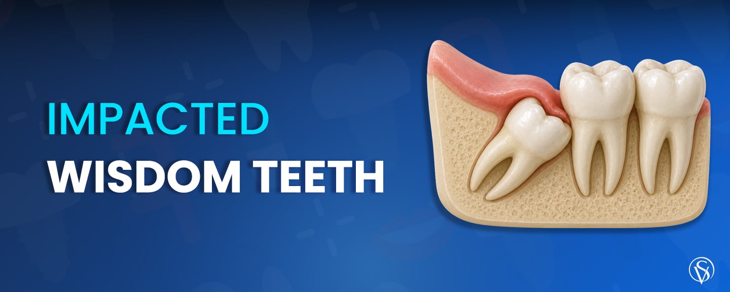
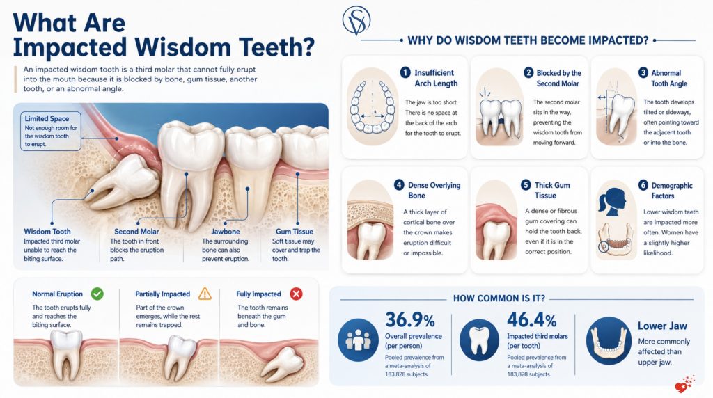
An impacted wisdom tooth is a third molar blocked from reaching a functional position in the bite, either by insufficient space at the back of the arch, by the tooth in front of it, or by a developmental angle pointing into bone rather than into the mouth. Wisdom teeth arrive last, between the ages of 17 and 24, and the lower jaw is affected more often than the upper. Impaction is common rather than exceptional, with a pooled worldwide prevalence of 36.9% per subject (Pinto et al., 2024).
Impaction is classified by angulation and by depth, and position predicts the damage a tooth can cause to the second molar beside it. Symptoms range from recurrent gum swelling and restricted jaw opening to nothing at all, so an asymptomatic tooth is not a healthy tooth. The decisive question is whether a given tooth has to come out, and the evidence does not support removing every one of them. Where surgery is indicated, its risks and the recovery timeline are predictable.
Key Points
- An impacted wisdom tooth is a third molar blocked from reaching a functional position in the bite by insufficient space, by the adjacent tooth, or by an abnormal developmental angle.
- Impaction affects 36.9% of people (95% CI 33.1–40.7%) across 98 studies and 183,828 subjects, and lower third molars are impacted more often than upper ones (Pinto et al., 2024).
- An asymptomatic tooth is not a disease-free tooth, because decay on the second molar, early bone loss, and cystic change are radiographic findings before they become painful ones (Ghaeminia et al., 2020).
- Decay on the adjacent second molar is the best-documented consequence at 29.89% (95% CI 21.05–38.74%), and the risk is highest in mesioangular teeth at Pell and Gregory level A (Revuelta-Cortés et al., 2025).
- Routine prophylactic removal of asymptomatic disease-free impacted wisdom teeth is not supported by current evidence, and watchful monitoring may be the more prudent strategy (Ghaeminia et al., 2020).
- Coronectomy limits inferior alveolar nerve injury to 0.59% of procedures, against transient injury of up to 16.66% and permanent injury of up to 3.63% after full surgical extraction (Póvoa et al., 2021; Abu-Mostafa et al., 2021).
What Are Impacted Wisdom Teeth?
An impacted wisdom tooth is a third molar prevented from reaching the occlusal plane by a physical obstruction. That obstruction takes one of three forms, which are a lack of space at the posterior end of the arch, the crown of the second molar sitting across the eruption path, or an abnormal developmental angle. Third molars are the only teeth that routinely fail to erupt, and third molar impaction is recognised as a developmental condition in international disease classification. Impacted third molars are identified during routine radiographic review in general dentistry, often before a patient notices anything.
The condition is far more prevalent than most patients expect. A meta-analysis of 98 studies covering 183,828 subjects found a pooled prevalence of 36.9% (95% CI 33.1–40.7%) per subject and 46.4% (95% CI 36.7–56.1%) per tooth, with the highest rates in Asia at 43.1% and the lowest in Europe at 24.5% (Pinto et al., 2024). That review also found a slightly higher likelihood in women, with an odds ratio of 1.173, and confirmed that mandibular impaction outnumbers maxillary impaction. An earlier meta-analysis of 49 studies and 83,484 individuals aged 17 and over reported a lower figure of 24.4% (95% CI 19.0–30.8%) (Carter & Worthington, 2016). The gap between 24.4% and 36.9% is methodological rather than biological, since studies differ in where they draw the line between a tooth that is merely late and a tooth that is blocked.
What Is the Difference Between an Impacted, a Partially Erupted, and an Unerupted Wisdom Tooth?
Three terms describe three different states and they are not interchangeable. Each state carries its own risk profile and its own management, so the distinction changes what happens next rather than only what the tooth is called. A tooth can be invisible in the mouth and blocked from erupting, or fully visible and still blocked. Five criteria separate the three states.
| Aspect | Unerupted | Partially erupted | Impacted |
| Position | Under the gum, still inside its expected developmental window | Crown through the gum but short of the biting plane | Blocked from ever reaching the biting plane |
| Visible in the mouth | No | Partly | From fully buried to partly visible, depending on depth |
| Diagnosis carried | None, the tooth is on schedule | Not a diagnosis on its own | A developmental condition requiring assessment |
| Main risk | None while the eruption window is open | Pericoronitis and distal caries, because the pocket cannot be cleaned | Cystic change, root resorption, and distal caries, weighted by depth and angle |
| Standard management | Review at the next routine appointment | Clinical assessment and radiograph | Radiographic assessment, then removal where disease is present or monitoring where it is not |
The categories are not fully exclusive, and this is where most patient confusion starts. Partial eruption describes what is visible, while impaction describes whether the tooth can ever reach function, so one tooth can be both partially erupted and impacted at the same time. That combination is the highest-risk version, because it pairs a permanent opening into the follicle with a tooth that will never complete its eruption. An unerupted third molar in a 19 year old and a partially erupted impacted tooth in the same patient are two different clinical situations requiring two separate plans.
Why Do Wisdom Teeth Become Impacted?
Impaction is a mechanical outcome, so its causes are the physical obstacles standing between a developing tooth and its place in the arch. Jaw growth finishes before the third molar finishes forming, which means the available space is already fixed while the tooth is still moving. Demographic factors shift the likelihood without explaining any individual case. Six recognised causes account for most impactions.
- Insufficient arch length: The jaw finishes growing before the third molar completes its root, leaving no space at the posterior end of the arch for the crown to emerge into.
- Obstruction by the second molar: The crown of the tooth in front sits directly across the eruption path, and the third molar meets it instead of the biting plane.
- Abnormal developmental angulation: The tooth forms tilted, and its long axis points into the adjacent tooth, into bone, or into the ramus rather than upward.
- Dense overlying bone: A thick layer of cortical bone over the crown resists eruption in a way that thinner bone does not.
- Thick or fibrous overlying soft tissue: A dense mucosal covering can hold back a tooth that is otherwise correctly angled and adequately spaced.
- Demographic factors: Women carry a slightly higher likelihood of impaction, with an odds ratio of 1.173, and lower third molars are impacted more frequently than upper ones (Pinto et al., 2024).
These causes are not mutually exclusive, and most impacted teeth combine two or three of them. A mesially tilted lower third molar in a short arch is a different clinical problem from a vertically positioned tooth held back only by soft tissue, even though both are recorded as impacted. This is why a diagnosis of impaction on its own says very little about what should happen next. What determines the plan is position, depth, and whether disease is present.
What Are the Types of Impacted Wisdom Teeth?
Impacted third molars are classified in two dimensions, and both appear in surgical planning notes. Winter’s classification records the angle of the impacted tooth relative to the long axis of the second molar. Pell and Gregory classification records how deeply the tooth sits in bone and how much of it is buried in the ramus of the mandible. Angulation is the dimension patients hear most about, and five patterns cover almost every case.
- Mesioangular impaction: The crown tilts forward toward the second molar, which is the most frequent pattern and the one most closely linked to decay on the neighbouring tooth.
- Distoangular impaction: The crown tilts backward away from the second molar and into the ramus, which makes surgical access more demanding.
- Vertical impaction: The tooth is correctly oriented but blocked from erupting fully by bone or soft tissue above it.
- Horizontal impaction: The tooth lies on its side with the crown facing directly into the roots of the second molar.
- Buccal or lingual impaction: The tooth is displaced toward the cheek or the tongue rather than tilted forward or backward, which is more common in the upper jaw.
Depth is recorded alongside angulation. Pell and Gregory levels A, B, and C describe a crown at, partly below, or entirely below the biting surface of the second molar, while classes I, II, and III describe how much of the tooth is covered by the ramus. Together the two systems produce a shorthand such as mesioangular level A class I, and that shorthand carries a specific risk profile. Both systems date to the 1930s and remain the language of surgical planning.
What Is the Difference Between Soft Tissue and Bony Impaction?
Soft tissue impaction means the crown of the tooth is covered only by gum, with no bone over it. Partial bony impaction means bone still covers part of the crown, and full bony impaction means the tooth is entirely encased. The distinction is separate from angulation, and a tooth can be correctly angled yet fully buried in bone.
This is the single variable that most affects how a removal is performed. A soft tissue impaction may need only an incision and elevation, while a full bony impaction requires bone removal and sectioning of the tooth. Post-operative swelling, restricted jaw opening, and healing time all scale with how much bone has to be removed.
Which Impaction Position Causes the Most Problems?
The mesioangular position causes the most problems. A meta-analysis of 13 studies covering 13,788 patients found that the mesioangular angulation was the position most frequently associated with distal caries in the adjacent second molar, at 43.37% (95% CI 33.03–53.70%), and that Pell and Gregory position A was associated with caries in 33.97% of cases (95% CI 24.06–43.89%) (Revuelta-Cortés et al., 2025).
The combination is what matters most. A mesioangulated tooth at level A carries the highest recorded risk of decay on the second molar, because the forward tilt creates a plaque trap against the neighbouring root surface at exactly the height where a toothbrush cannot reach. Knowing this changes the monitoring interval rather than automatically triggering surgery. A vertically impacted tooth deep in bone and a mesioangular tooth at level A are not equivalent findings and do not warrant equivalent follow-up.
What Are the Symptoms of an Impacted Wisdom Tooth?
Symptoms of an impacted wisdom tooth come from two sources, which are inflammation of the gum around a partially erupted crown and pressure or infection deeper in the jaw. Because the same tooth can be silent for years and then flare within days, symptoms tend to arrive in episodes rather than steadily. Recognising the early pattern shortens the time to treatment. Seven presentations account for most cases.
- Pain at the back of the jaw: A dull ache behind the last visible tooth that comes and goes over weeks, sometimes triggered by chewing.
- Swollen or bleeding gum over the tooth: Inflamed tissue covering a partially erupted crown, which bleeds on brushing and feels tender to the tongue.
- Bad taste or persistent bad breath: Food debris and bacteria held in the pocket behind the second molar, producing a taste that returns after brushing.
- Difficulty opening the mouth: Restricted opening caused by inflammation spreading into the muscles around the joint.
- Pain on biting: Discomfort when the opposing tooth contacts inflamed tissue over the impacted crown.
- Tender lymph nodes under the jaw: Swollen nodes on the affected side, indicating that infection has moved beyond the local tissue.
- Food trapping behind the second molar: A pocket that catches food at every meal and cannot be cleaned with a brush or floss.
Any one of these presentations is a reason for a clinical examination and a radiograph rather than a wait-and-see approach. Episodes of pericoronitis tend to recur, and each recurrence carries more weight in the decision about removal than the severity of a single episode. Recording how often symptoms return is more useful than recording how badly they hurt. That history is what a surgeon uses when weighing removal against continued monitoring.
Warning: Spreading swelling of the face or neck, fever, difficulty swallowing or breathing, or an inability to open the mouth are signs that infection has moved beyond the tooth. These require same-day clinical assessment rather than a routine appointment.
Can an Impacted Wisdom Tooth Be Painless?
Yes. An impacted wisdom tooth can be entirely painless and still be causing damage, which is why absence of symptoms is not evidence of absence of disease. The clinical definition of an asymptomatic disease-free impacted wisdom tooth requires both the absence of symptoms and the absence of radiographic evidence of local disease, and the two conditions are assessed separately (Ghaeminia et al., 2020).
The practical consequence is that decay on the distal surface of the second molar, early periodontal breakdown, and cystic change in the follicle are all radiographic findings before they are painful findings. A patient with no complaints can have a second molar already losing tooth structure at the contact point. This is the reason a retained third molar is followed with periodic radiographs rather than left until something hurts.
What Problems Can an Impacted Wisdom Tooth Cause?
The problems caused by an impacted wisdom tooth are almost all problems for the tooth beside it or the tissue around it, rather than for the impacted tooth itself. Infection risk concentrates in partially erupted teeth, while resorption and cystic change concentrate in fully buried ones. The strength of evidence differs across the list, and it is worth saying which findings are well established and which are not. Seven recognised complications are documented.
- Pericoronitis: Acute inflammation of the gum around a partially erupted crown, which recurs and is the most frequent reason for removal.
- Distal caries in the second molar: Decay on the back surface of the adjacent tooth at the point where the impacted crown creates an uncleansable contact.
- Periodontitis around the second molar: Bone and attachment loss on the distal aspect of the neighbouring tooth, supported by very low certainty evidence over the long term (Ghaeminia et al., 2020).
- Caries in the impacted tooth itself: Decay in a partially exposed crown that cannot be restored predictably because of access.
- External root resorption: Loss of root structure on the second molar where an impacted crown presses directly against it.
- Cystic or tumoural change in the follicle: Expansion of the tissue sac around a buried tooth, which is uncommon but consequential when it occurs.
- Recurrent soft tissue trauma: Repeated cheek or tongue biting caused by a tooth displaced toward the buccal or lingual side.
Not every impacted tooth develops any of these, and the honest position is that the risk is real but not universal. The Cochrane assessment of periodontitis risk in adjacent second molars is rated very low certainty, which means it is a signal rather than a settled fact (Ghaeminia et al., 2020). Decay on the second molar, by contrast, has consistent prevalence data behind it. Where gum disease around a retained third molar is already established, treatment falls to periodontics alongside the decision about the tooth.
Can an Impacted Wisdom Tooth Damage the Neighbouring Tooth?
Yes. Damage to the adjacent second molar is the best-documented consequence of retaining an impacted third molar. The overall prevalence of distal caries in mandibular second molars next to an impacted mandibular third molar is 29.89% (95% CI 21.05–38.74%), pooled across 13 studies and 13,788 patients (Revuelta-Cortés et al., 2025).
Reported figures vary widely depending on which population is examined. A systematic review and meta-analysis of patients referred for third molar assessment found distal surface caries prevalence ranging from 4.5% to 52% across 11,452 second molars (Toedtling et al., 2019). The high end of that range comes from studies using cone beam CT, which detects lesions a panoramic radiograph misses. What the numbers agree on is direction rather than magnitude, since the contact point between an impacted crown and a second molar root is a site that cannot be cleaned. Once a lesion is established there, the impacted tooth and the decay are two separate problems requiring two separate treatments, and the restoration of a tooth cavity on that surface is technically demanding because of access.
Do Impacted Wisdom Teeth Cause Crowding of the Front Teeth?
No. The belief that impacted lower wisdom teeth push the front teeth out of alignment is not supported by trial evidence. A single randomised trial in adolescents comparing removal of impacted mandibular wisdom teeth with retention found no evidence of an effect on late lower incisor crowding over 5 years, and that trial was assessed as being at high risk of bias (Ghaeminia et al., 2020).
Preventing late lower incisor crowding has been offered as a justification for prophylactic removal for decades, and it is the weakest of the arguments made. Late crowding of the lower front teeth occurs in patients whose third molars were removed years earlier and in patients who never developed third molars at all. Where alignment is the concern, the causes and treatment of crooked teeth are assessed on their own terms rather than through the third molars.
How Are Impacted Wisdom Teeth Diagnosed?
Diagnosis of an impacted wisdom tooth is radiographic, because the position, depth, and angle of the tooth cannot be determined by looking in the mouth. A clinical examination establishes whether the tooth is partially erupted, whether the gum around it is inflamed, and how far the patient can open. Imaging then establishes everything that determines the plan. Three assessments make up a complete diagnosis.
- Clinical examination: Inspection of the eruption state, probing of the pocket behind the second molar, assessment of jaw opening, and palpation of the lymph nodes.
- Panoramic radiograph: The standard image for recording angulation, Pell and Gregory level and class, root formation, and the relationship of the roots to the inferior alveolar canal.
- Cone beam CT: A three-dimensional image requested where a panoramic radiograph suggests the roots sit close to the inferior alveolar canal and the surgical approach depends on that relationship.
Cone beam CT is not part of a routine assessment and is requested for a specific reason. Radiographic signs on a panoramic image that raise the question include interruption of the white line of the canal, darkening of the root where it crosses the canal, and deflection of the canal itself. Where those signs appear, three-dimensional imaging changes the surgical decision rather than simply confirming it. This is also the point at which coronectomy enters the discussion as an alternative to full removal.
Should Every Impacted Wisdom Tooth Be Removed?
No. Not every impacted wisdom tooth needs to be removed, and the routine removal of asymptomatic disease-free impacted third molars is not supported by current evidence. A Cochrane review found insufficient evidence to support or refute routine prophylactic removal in adults, and concluded that watchful monitoring of asymptomatic third molars may be the more prudent strategy (Ghaeminia et al., 2020).
Two considerations pull in opposite directions, and both are legitimate. Retaining a tooth carries the risk that disease develops in the adjacent second molar over years. Removing a tooth carries immediate surgical risk, and that risk is not constant across a lifetime, since the Cochrane review notes that the frequency and severity of post-operative complications, pain, and discomfort increase when removal is performed in older patients (Ghaeminia et al., 2020). Neither consideration settles an individual case on its own. What settles it is whether disease is present, what the position of the tooth predicts, and what the patient’s own risk of decay and gum disease looks like.
Clinic Note: An assessment appointment for an impacted wisdom tooth should produce four recorded findings, which are the angulation and Pell and Gregory classification, the presence or absence of disease in the tooth and the second molar, the relationship of the roots to the inferior alveolar canal, and a documented review interval where the tooth is retained. A decision to monitor is a clinical decision with a follow-up date attached, not a decision to take no action.
When Is It Recommended to Have an Impacted Wisdom Tooth Extracted?
Removal is recommended when disease is present or when a planned treatment makes retention untenable. The presence of pathology, rather than the presence of impaction, is what moves a tooth from monitoring to surgery. This is a shorter list than patients expect. Seven indications cover the recognised reasons for removal.
- Recurrent pericoronitis: Repeated episodes of acute inflammation around a partially erupted crown, where each recurrence carries more weight than the severity of any single episode.
- Caries in the impacted tooth: Decay in a crown that cannot be restored predictably because access is inadequate.
- Caries in the adjacent second molar: Decay on the distal surface of the neighbouring tooth caused by the contact with the impacted crown.
- Periodontal disease at the distal of the second molar: Established attachment and bone loss on the back surface of the adjacent tooth.
- Cystic or tumoural change: Radiographic expansion of the follicle around a buried tooth.
- External root resorption: Loss of root structure on the second molar where the impacted crown presses against it.
- Planned orthognathic surgery or radiotherapy to the field: Preparation for jaw surgery or for radiotherapy involving the area, both of which are recognised justifications for removing an otherwise asymptomatic tooth (Ghaeminia et al., 2020).
An indication on this list is a reason to discuss removal rather than an instruction to proceed. Position, depth, proximity to the inferior alveolar canal, and the patient’s age and general health all modify what the safest course is. In some cases the appropriate answer is coronectomy rather than full removal. In others the appropriate answer is to treat the second molar and continue monitoring the third.
What Does Watchful Monitoring Involve?
Watchful monitoring means active clinical and radiographic follow-up of a retained tooth on a defined schedule. It involves a clinical examination of the gum and pocket behind the second molar, a review of any symptom episodes since the last visit, and a periodic radiograph to check the distal surface of the second molar and the follicle around the impacted crown. The interval is set according to position and risk rather than applied uniformly.
Monitoring is not the same as leaving a tooth alone. A mesioangular tooth at level A in a patient with a history of decay warrants closer follow-up than a deeply buried vertical tooth in a patient with no caries history. The purpose of the schedule is to detect a distal lesion or early bone loss while it is still small, at which point the decision to remove the tooth is made on evidence rather than on prediction. Patients on a monitoring schedule also need to know which symptoms bring the next appointment forward.
How Is an Impacted Wisdom Tooth Removed?
Removing an impacted wisdom tooth is a surgical procedure rather than a simple extraction, because access has to be created before the tooth can be delivered. The extent of the surgery scales with how much bone covers the crown and how the tooth is angled. A soft tissue impaction and a full bony impaction sit at opposite ends of that scale. Seven stages make up the procedure, and the middle stages are omitted where they are not needed.
- Assessment and imaging: Confirmation of angulation, depth, and the relationship of the roots to the inferior alveolar canal, with three-dimensional imaging where that relationship is unclear.
- Local anaesthesia: Infiltration and block anaesthesia to the surgical field, checked before any incision is made.
- Flap raising: An incision in the gum and reflection of the soft tissue to expose the bone and the crown.
- Bone removal: Removal of the bone covering the crown, carried out with irrigation, and omitted entirely in a soft tissue impaction.
- Sectioning of the tooth: Division of the crown and roots so that each fragment can be delivered along a path that does not damage the second molar.
- Delivery: Elevation and removal of the fragments, with the socket checked for retained tooth structure.
- Irrigation and closure: Debridement of the socket, irrigation, and suturing of the flap back into position.
The number of stages actually used is what separates a short procedure from a long one, and it is determined by the radiograph rather than by the surgeon’s preference. This is also why post-operative swelling and restricted opening vary so widely between patients who both had a wisdom tooth removed. A tooth requiring bone removal and sectioning produces a different recovery from one lifted out from under soft tissue. The general principles of tooth extraction apply throughout, with the additional steps required by impaction layered on top.
Is an Impacted Wisdom Tooth Removed Under Local or General Anaesthesia?
Impacted wisdom teeth are removed under local anaesthesia in the great majority of cases. Local anaesthesia gives complete control of the surgical field, allows the patient to report unexpected sensation in the lip or tongue during the procedure, and avoids the additional risk that comes with general anaesthesia.
Sedation is considered in specific circumstances rather than as a default. Marked dental anxiety, removal of several impacted teeth in one appointment, deeply buried teeth requiring extended surgical time, and a strong gag reflex are the situations in which sedation dentistry changes the plan. General anaesthesia is reserved for cases where sedation is inadequate or where medical factors require it.
What Is a Coronectomy?
A coronectomy is the intentional removal of the crown of a lower third molar with the roots deliberately left in place. The tooth is sectioned at the cemento-enamel junction, the crown is removed, and the root surface is smoothed and left below the level of the bone. The purpose is narrow, which is to avoid direct trauma to the inferior alveolar nerve in teeth whose roots sit against the canal.
The recorded complication profile is low. Across 16 papers analysing 2,176 coronectomies, inferior alveolar nerve injury occurred in 0.59% of procedures, lingual nerve injury in 0.22%, infection in 3.95%, dry socket in 1.12%, unintended removal of the root in 5.28%, and reintervention in 1.13% (Póvoa et al., 2021). Movement of the retained roots is expected rather than exceptional, with migration of 2.33 to 3.43 mm recorded at 6 months and stabilising by 12 months (Abu-Mostafa et al., 2021). Root migration moves the fragment away from the nerve, which is why a later removal, if it becomes necessary, carries a lower risk than the original surgery would have.
What Is the Difference Between Coronectomy and Full Surgical Extraction?
The two procedures differ in what is left behind and in the risk each carries to the inferior alveolar nerve. The comparison applies only to lower third molars whose roots lie against the canal, since that is the only situation in which coronectomy is indicated.
| Aspect | Coronectomy | Full surgical extraction |
| What is removed | Crown only, roots retained below bone level | Entire tooth including roots |
| Transient nerve injury | 0–2.20% in successful cases, 0–8% in failed cases | 0–16.66% |
| Permanent nerve injury | Not reported in the included trials | 0–3.63% |
| Root migration | 2.33–3.43 mm at 6 months, stabilising by 12 months | Not applicable |
| Reintervention | 1.13% across 2,176 procedures | Not applicable |
| Candidacy | Lower third molars with roots against the inferior alveolar canal | All other impacted third molars |
Figures for nerve injury are drawn from a systematic review of trials comparing the two procedures in patients at high risk (Abu-Mostafa et al., 2021), and figures for reintervention and migration from a review of 2,176 coronectomies (Póvoa et al., 2021). The trade-off is straightforward, since coronectomy substantially reduces nerve injury risk while accepting a small chance of needing a second procedure. It is not an alternative for routine impactions, and it is not performed on upper third molars. Where the roots are clear of the canal, full removal remains the appropriate procedure.
What Are the Risks of Removing an Impacted Wisdom Tooth?
Removing an impacted wisdom tooth carries surgical risks, and their frequency depends on the depth of the tooth, its proximity to the inferior alveolar canal, and the age of the patient. Most complications are temporary and resolve within two weeks. A smaller number are persistent, and nerve injury is the one that matters most in consent. Nine complications are documented.
- Inferior alveolar nerve injury: Altered or reduced sensation in the lower lip and chin, reported as transient in 0–16.66% of extractions and permanent in 0–3.63% across the included trials (Abu-Mostafa et al., 2021).
- Lingual nerve injury: Altered sensation or taste on one side of the tongue, arising from retraction or instrumentation on the lingual side.
- Dry socket: Breakdown or loss of the blood clot with exposure of bone, producing severe pain that begins 2 to 4 days after surgery.
- Surgical site infection: Delayed swelling, discharge, and pain, recorded at 3.95% after coronectomy in a review of 2,176 procedures (Póvoa et al., 2021).
- Trismus: Restricted mouth opening caused by inflammation in the muscles of mastication, which resolves as the swelling settles.
- Swelling and pain: Expected post-operative inflammation that peaks in the first 48 hours rather than immediately after surgery.
- Prolonged bleeding: Oozing from the socket that continues beyond the first few hours, more likely where anticoagulant medication is involved.
- Root fracture: Separation of a root tip during delivery, which is sometimes left in place deliberately when retrieval would risk the nerve.
- Damage to the adjacent second molar: Loosening, fracture of a restoration, or injury to the root surface of the neighbouring tooth.
Consent for this procedure is meaningful only if nerve injury is discussed explicitly, with the individual radiographic risk rather than the average figure. A tooth whose roots are clearly separate from the canal presents a different conversation from one where the canal is deflected around the root. Smoking, anticoagulant medication, poor oral hygiene, and existing pericoronitis all raise complication rates independently of the tooth itself. These factors are modifiable before surgery in a way that the position of the tooth is not.
Warning: Pain that intensifies 2 to 4 days after surgery, accompanied by a bad odour and a socket that looks empty, points to dry socket rather than normal healing. This needs clinical review and socket dressing rather than higher doses of painkillers. Numbness of the lip, chin, or tongue that has not started to improve within a few weeks also needs review rather than continued waiting.
Does Age Increase the Risk of Complications?
Yes. Age increases both the frequency and the severity of complications after removal of an impacted wisdom tooth. The Cochrane review states that when surgical removal is performed in older people, the risk of post-operative complications, pain, and discomfort is increased (Ghaeminia et al., 2020).
Two mechanisms explain the pattern. Bone becomes denser with age, so more bone has to be removed to deliver the same tooth, and root formation completes, which removes the mechanical advantage available when roots are still short. The consequence is a genuine tension in the evidence, since the argument for early removal rests on lower surgical risk while the argument against it rests on the absence of proof that removal is needed at all. That tension is unresolved, and any article claiming otherwise is overstating the evidence.
What Is the Recovery Like After Impacted Wisdom Tooth Removal?
Recovery after removal of an impacted wisdom tooth follows a predictable sequence, with the difficult period concentrated in the first week. Swelling peaks later than patients expect, so day two often feels worse than the day of surgery. Healing of the soft tissue is complete well before the socket has filled with bone. Three phases describe the process.
- First 24 hours: Clot formation and bleeding control. Pressure on the gauze pack, no rinsing, no smoking, and no drinking through a straw, since all of these disturb the clot.
- Days 2 to 7: Peak swelling and restricted opening at around 48 hours, then steady improvement. Careful brushing away from the socket, warm saline rinses from day two, and soft foods throughout.
- Weeks 2 to 4: Resolution of swelling and opening, closure of the soft tissue, and removal of sutures where non-resorbable material was used. Bone fill of the socket continues for months after the surface has healed.
Deviation from this sequence is the signal to seek review. Pain that decreases and then sharply increases, swelling that grows after day three rather than shrinking, and fever at any stage are all outside the normal pattern. Numbness of the lip or tongue immediately after surgery is common and resolves as the anaesthetic wears off, but numbness persisting the following day needs to be recorded and followed. Recovery from a soft tissue impaction is considerably shorter than recovery from a deeply buried tooth requiring bone removal.
Tips for Patients: Apply a cold compress to the outside of the face in 20 minute cycles during the first 24 hours, then switch to warm compresses. Sleep with the head elevated on an extra pillow for the first two nights. Start warm saline rinses on day two rather than day one, and brush the rest of the mouth normally while avoiding the socket itself. Avoid smoking, alcohol, and straws for at least 7 days, since all three disturb the clot.
How Long Does Swelling Last After Impacted Wisdom Tooth Removal?
Swelling after removal of an impacted wisdom tooth peaks at around 48 hours and settles substantially by day 7. Those two points, 48 hours and the seventh post-operative day, are the standard measurement intervals used in surgical trials, which is why they describe the curve accurately.
Restricted mouth opening follows the same curve slightly behind the swelling, and full opening returns over 2 to 3 weeks. The amount of swelling correlates with the amount of bone removed and the duration of surgery rather than with the patient’s pain tolerance. A patient whose tooth needed sectioning and bone removal can expect visible facial swelling, while a soft tissue impaction may produce almost none.
What Should You Eat and Avoid After Impacted Wisdom Tooth Surgery?
Diet after removal of an impacted wisdom tooth is soft, cool, and swallowed rather than chewed for the first few days. The purpose is to protect the clot in the socket rather than to reduce pain. Nutrition still matters during the first week, so soft does not mean minimal. Four groups cover what to eat and what to leave alone.
- Soft, Swallowable Foods (Day 1 Onward): Yoghurt, smooth soups at room temperature, mashed potato, scrambled eggs, blended fruit, and porridge are safe from the first day.
- Soft, Chewable Foods (From Day 3): Soft pasta, flaked fish, soft rice, well-cooked vegetables, and minced meat in sauce can be introduced once the socket is less sensitive.
- Hard or Sharp Foods (Avoid for 7 Days): Crunchy food such as crisps and nuts, and seeds and grains that lodge in the socket, can dislodge the clot or become trapped in the wound.
- Hot or Irritant Foods and Drinks (Avoid for 7 Days): Hot liquids, spicy food, acidic drinks, and alcohol increase blood flow to the socket or irritate the healing tissue.
Returning to a normal diet is gradual and led by comfort rather than by the calendar. Most patients are chewing normally on the operated side within 2 weeks. The socket continues to trap food for several weeks after it stops being painful, so gentle irrigation with a syringe, where the clinic provides one, is more effective than brushing at that stage. Persistent food trapping beyond a month is worth reviewing.
Warning: Diet is only half of clot protection, and the other half is behaviour. Drinking through a straw, smoking, vigorous rinsing, and chewing on the surgical side all create suction or pressure that can pull the clot out of the socket, and all four should be avoided for 7 days. Clot loss produces dry socket, with pain beginning 2 to 4 days after surgery.
How Much Does Impacted Wisdom Tooth Removal Cost?
The cost of removing an impacted wisdom tooth is driven by impaction depth rather than by the tooth being a wisdom tooth. A soft tissue impaction and a full bony impaction are billed as different procedures in every pricing system, and the gap between them is several times the base fee. Anaesthesia, imaging, and the number of teeth treated in one session sit on top of that. The figures here are converted to euro for comparison.
| Country | Single impacted tooth (Average Cost) | All four impacted (Average Cost) |
| Turkey | €150–€400 | €185–€465 |
| United Kingdom | €400–€920 | €690–€1,380 |
| United States | €275–€1,010 | €2,200–€3,830 |
The single-tooth and all-four columns do not scale, and the reason is worth stating plainly. Turkish clinics price four teeth as one surgical session, so the all-four figure lands close to the single-tooth figure rather than four times it. In the United States each tooth is billed under its own impaction code, which is why four impacted teeth reach €2,200 or more. Anyone comparing quotes across countries should confirm whether imaging, anaesthesia, and follow-up are inside the figure, since a full breakdown of wisdom teeth removal cost shows those components excluded from most headline prices.
Which Factors Affect the Cost of Impacted Wisdom Tooth Removal?
Two teeth in the same mouth can carry very different fees, and the difference is recorded on the radiograph before any quote is given. Depth and angulation determine how much surgical time is needed, and surgical time is what pricing tracks. Six factors account for almost all of the variation.
- Impaction depth: Soft tissue, partial bony, and full bony impaction are priced as separate procedures, with full bony impaction at the top of the range.
- Angulation: Distoangular and horizontal teeth need more sectioning and more access than vertical teeth at the same depth.
- Anaesthesia type: Local anaesthesia is inside the base fee, while IV sedation adds €230–€460 and general anaesthesia more again.
- Imaging: A panoramic radiograph is standard, and cone beam CT is charged separately where the roots sit against the inferior alveolar canal.
- Number of teeth per session: Treating several teeth in one appointment lowers the per-tooth figure, since theatre time and anaesthesia are charged once.
- Procedure chosen: Coronectomy and full surgical extraction are priced differently, and coronectomy carries a small chance of a second procedure later.
Insurance changes the number a patient actually pays rather than the fee itself. Removal for a documented clinical indication is treated differently from removal of an asymptomatic tooth in most systems, and asymptomatic removal is often excluded entirely. This matters because the evidence does not support routine prophylactic removal, so a patient requesting it on a preventive basis is likely to be paying the full figure. Getting the indication recorded before the quote is the practical step.
FAQ
The surgical time depends almost entirely on how much bone covers the crown and whether the tooth needs sectioning. A soft tissue impaction can be delivered in a few minutes, while a deeply buried distoangular lower tooth requiring bone removal and sectioning takes considerably longer. The appointment itself is longer than the surgery, since it includes anaesthesia, waiting for it to take effect, and post-operative instructions.
Yes. All four can be removed in a single appointment, and this is often done where sedation or general anaesthesia is already planned. The trade-off is a single recovery period that is more uncomfortable than any individual quadrant would be, with swelling on both sides and greater difficulty eating. Removing one side at a time keeps one side of the mouth functional throughout, which some patients prefer.
Antibiotics are not required routinely after every removal. Evidence indicates that antibiotic use reduces the incidence of infection and dry socket compared with no prescription, but the decision balances that reduction against antibiotic resistance and side effects. Prescription is more likely where the tooth was infected before surgery, where surgery was extended, or where the patient has a relevant medical condition.
An impacted wisdom tooth cannot be made to erupt, so the alternatives to removal are monitoring and management of symptoms rather than a different way of resolving the impaction. An acute episode of pericoronitis is treated with irrigation, debridement of the pocket, and occasionally antibiotics, which settles the episode without addressing the cause. Where no disease is present, monitoring on a defined schedule is a recognised strategy (Ghaeminia et al., 2020).
Flying is generally safe once bleeding has stopped and the clot is stable, which for most patients is after 48 to 72 hours. The greater consideration is access to care rather than the flight itself, since dry socket and infection both present 2 to 4 days after surgery, exactly when a traveller may be furthest from the treating clinic. Long-haul travel within the first week leaves little margin if a complication develops.
Yes. Pain from an impacted lower wisdom tooth is frequently felt in the ear, the angle of the jaw, or the temple rather than at the tooth itself, because the nerves supplying the area share pathways with those structures. Restricted opening from inflammation in the chewing muscles adds a separate ache around the joint and temple. Referred pain of this kind does not indicate that the infection has spread to the ear.
Migration of the retained roots is expected rather than a complication, with movement of 2.33 to 3.43 mm recorded at 6 months and stabilising by 12 months (Abu-Mostafa et al., 2021). Because the roots move away from the inferior alveolar canal, a later removal carries a lower nerve injury risk than the original surgery would have. Reintervention was needed in 1.13% of 2,176 coronectomies (Póvoa et al., 2021), and most retained roots require no further treatment.
Abu-Mostafa, N., AlRejaie, L. M., Almutairi, F. A., Alajaji, R. A., Alkodair, M. M., & Alzahem, N. A. (2021). Evaluation of the outcomes of coronectomy procedure versus surgical extraction of lower third molars which have a high risk for inferior alveolar nerve injury: A systematic review. International Journal of Dentistry, 2021, 9161606.
Carter, K., & Worthington, S. (2016). Predictors of third molar impaction: A systematic review and meta-analysis. Journal of Dental Research, 95(3), 267–276.
Ghaeminia, H., Nienhuijs, M. E. L., Toedtling, V., Perry, J., Tummers, M., Hoppenreijs, T. J. M., Van der Sanden, W. J. M., & Mettes, T. G. (2020). Surgical removal versus retention for the management of asymptomatic disease-free impacted wisdom teeth. Cochrane Database of Systematic Reviews, 2020(5), CD003879.
Pinto, A. C., Francisco, H., Marques, D., Martins, J. N. R., & Caramês, J. (2024). Worldwide prevalence and demographic predictors of impacted third molars: Systematic review with meta-analysis. Journal of Clinical Medicine, 13(24), 7533.
Póvoa, R. C. S., Mourão, C. F. A. B., Geremias, T. C., Sacco, R., Guimarães, L. S., Montemezzi, P., Cardarelli, A., Moraschini, V., Calasans-Maia, M. D., & Louro, R. S. (2021). Is coronectomy a feasible and safe procedure to avoid inferior alveolar nerve injury during third molar extractions? A systematic review. Healthcare, 9(6), 750.
Revuelta-Cortés, P., Cortés-Bretón Brinkmann, J., Argandoña-Flores, M., Pérez-González, F., Bazal-Bonelli, S., Madrigal Martínez-Pereda, C., & Meniz-García, C. (2025). Prevalence of distal caries in second molar associated with impacted mandibular third molar and the position and level of impaction: A systematic review and meta-analysis. Clinical Oral Investigations, 29(83).
Toedtling, V., Devlin, H., Tickle, M., & O’Malley, L. (2019). Prevalence of distal surface caries in the second molar among referrals for assessment of third molars: A systematic review and meta-analysis. British Journal of Oral and Maxillofacial Surgery, 57(6), 505–514.