
Remineralizing teeth is the redeposition of calcium, phosphate and fluoride ions into enamel that acid has stripped of mineral. It reverses early damage while the enamel surface is still unbroken, and it cannot repair a lesion that has already collapsed into a cavity. Enamel is around 96% mineral by weight and holds no living cells, so it does not heal the way skin or bone heals.
Enamel starts dissolving below pH 5.5, and plaque needs 20 to 60 minutes to return to neutral after each acid exposure, so how often you eat matters more than how much. Fluoride toothpaste at 1450 ppm is the strongest home measure, and hydroxyapatite paste matched it in an 18-month randomised trial. Roughly half of an early white spot lesion remineralizes within 6 months. Long-term follow-up shows about a third of these lesions become undetectable, while the rest fade without disappearing. Charcoal powders and homemade mineral rinses achieve none of this.
Key Points
- Remineralization restores mineral to enamel that is still intact, and it cannot close a cavity or rebuild lost enamel thickness.
- Enamel dissolves below pH 5.5 and plaque needs 20 to 60 minutes to recover after each acid exposure, so exposure frequency matters more than quantity.
- Fluoride toothpaste at 1450 ppm is the most effective home measure, and spitting without rinsing preserves its effect for the hours that follow.
- Hydroxyapatite paste matched 1450 ppm fluoride in an 18-month randomised trial, which makes it the one evidence-backed option for patients avoiding fluoride.
- About half of an early white spot lesion remineralizes within 6 months, with the fastest change in the first 6 weeks.
- Long-term follow-up shows roughly 37.5% of these lesions become undetectable, while the rest fade without disappearing completely.
What Is Remineralizing Teeth?
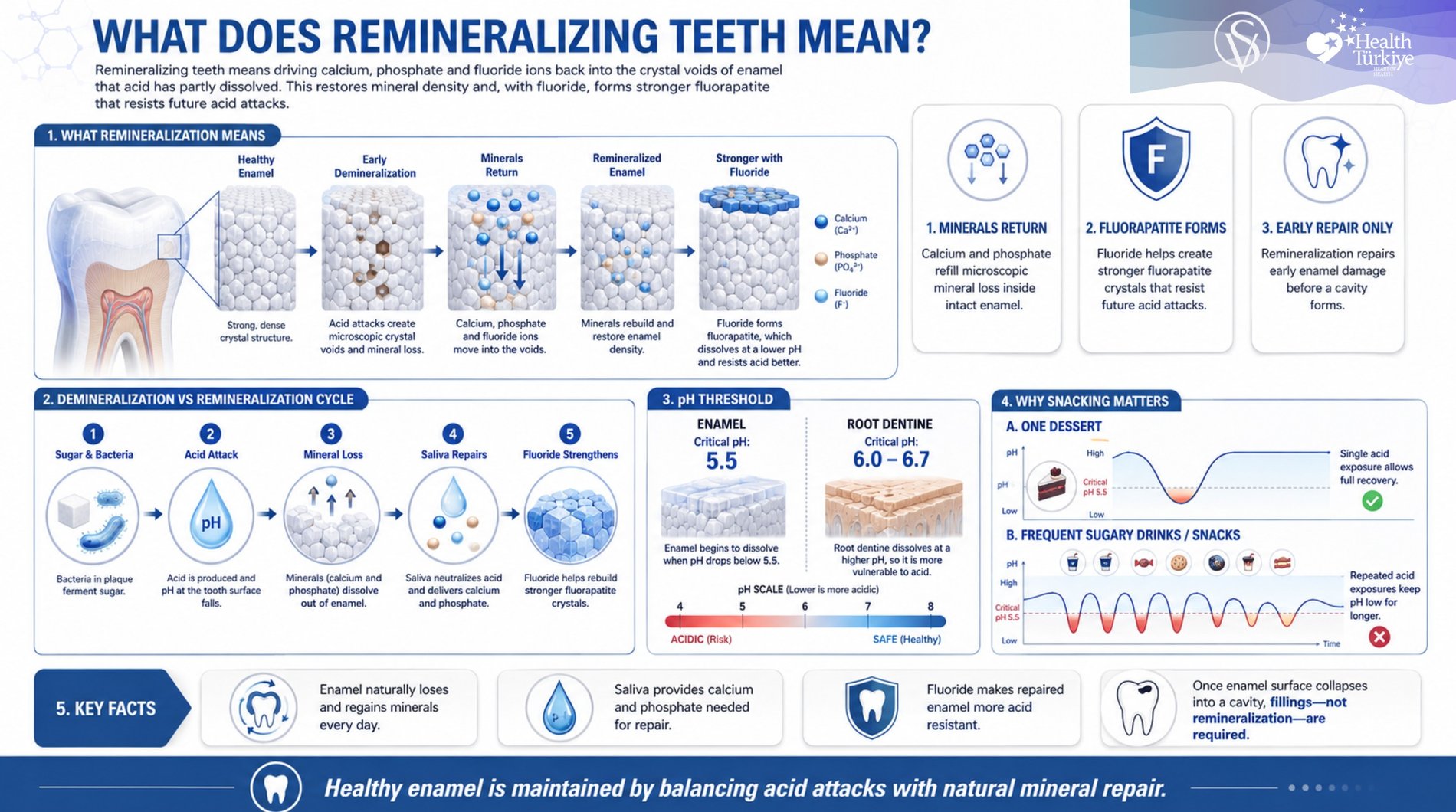
Remineralizing teeth is driving calcium, phosphate and fluoride ions back into the crystal voids of enamel that acid has partly dissolved. The process restores mineral density inside the enamel structure that is still present, and where fluoride is available it forms fluorapatite, a crystal that dissolves at a lower pH than the original hydroxyapatite and resists the next acid attack.
Enamel loses and regains mineral continuously through the day. Loss dominates during each acid exposure. Gain dominates in the hours between, as long as saliva flow is adequate and calcium and phosphate are available at the surface. A lesion only progresses when that balance stays negative long enough for the outer surface to break down.
Remineralisation is not enamel regrowth. Once the surface breaks down, the lesion becomes a tooth cavity and needs restorative treatment rather than mineral therapy. A treated white spot that becomes glossy and hard has responded successfully even when it stays visible.
What Causes Enamel to Lose Minerals?
Enamel loses mineral whenever acid reaches the tooth surface faster than saliva can neutralise it. Six conditions account for most mineral loss seen in practice:
- Plaque acid: Bacteria in undisturbed plaque convert sugars and cooked starches into organic acids that sit directly against the enamel surface.
- Dietary acid: Carbonated drinks, fruit juice, citrus, vinegar-based dressings and pickled foods drop the oral pH without any bacterial involvement.
- Gastric reflux: Stomach acid entering the mouth reaches pH 1.5 to 3.5 and attacks the inner surfaces of the upper teeth first, often overnight and without the patient noticing.
- Reduced saliva flow: Antihistamines, antidepressants, blood pressure medication, radiotherapy and mouth breathing all cut the buffering and mineral supply that repair depends on.
- Snacking and sipping frequency: Six small exposures cause more cumulative mineral loss than one larger meal, because each one restarts the pH recovery period.
- Fixed orthodontic appliances: Brackets and bands create plaque traps, and one prevalence study recorded white spot lesions in 46.6% of patients after 12 months of fixed treatment against 11.9% before treatment began.
Acid that reaches the tooth from food, drink or the stomach rather than from plaque bacteria produces dental erosion, which follows a different pattern of surface loss and needs a different prevention plan.
How Does Tooth Remineralization Work?
Tooth remineralization works through an acid cycle, plaque bacteria ferment carbohydrate into acid, pH at the tooth surface falls, mineral dissolves out of the enamel, then saliva neutralises the acid and delivers calcium and phosphate back into the softened surface.
Enamel begins to dissolve below pH 5.5. Exposed dentine and root surfaces dissolve earlier, from around pH 6.0 to 6.7, which is why sensitivity at the gum line responds to a milder acid load than a lesion on the biting surface. After one sugar exposure, plaque pH falls within 2 to 5 minutes and needs 20 to 60 minutes to recover. Each separate exposure restarts that clock. Ten sugared teas spread across a working day hold the tooth surface below the critical pH for far longer than one dessert eaten in five minutes, which is the mechanism behind the link between diet frequency and stronger teeth.
Fluoride changes the chemistry of the repair rather than only its speed. Ions deposited in the presence of fluoride form fluorapatite, which needs a pH below roughly 4.5 to dissolve. Repeated low-dose exposure from toothpaste produces this effect more reliably than occasional high-dose exposure.
How Reversible Is Enamel Damage?
Enamel damage is reversible only while the outer surface layer remains unbroken. Surface integrity is the dividing line, not lesion size or how dark the area looks. Reversibility falls into five levels.
- Fully reversible: Chalky white or matte lesions with an intact surface, scored ICDAS 1 or 2, regain mineral without any drilling.
- Arrestable but not erasable: Deeper enamel lesions stop progressing and harden under treatment, though the visible mark often remains for years.
- Partially treatable: Exposed dentine and root surfaces can be hardened and desensitised, while the tissue lost before treatment does not return.
- Not reversible: Lesions with a broken surface, scored ICDAS 3 and above, have no crystal framework left for mineral to enter and require restoration.
- Outside mineral therapy entirely: Enamel thickness lost to erosion, abrasion or grinding needs restorative build-up, because remineralisation adds density and not volume.
Patients arriving with a cavitated lesion are told directly that mineral therapy has no role for that tooth, and treatment moves to tooth fillings while prevention continues on the remaining teeth.
The evidence-based dividing line is set by the American Dental Association’s 2018 clinical practice guideline on nonrestorative treatments. Noncavitated lesions are managed with sealants plus 5% sodium fluoride varnish applied every 3 to 6 months. Cavitated coronal lesions are arrested with 38% silver diamine fluoride applied twice yearly or restored. Root surface lesions, cavitated or not, are managed with 5,000 ppm fluoride paste used daily at home.
Can Lost Enamel Grow Back?
No. Lost enamel cannot grow back, because the ameloblast cells that build enamel are lost when the tooth erupts and the body has no mechanism to produce more.
Remineralization refills mineral inside the crystal structure that survives. It does not add thickness, rebuild a worn cusp or close a cavity. Laboratory and early clinical work continues, including a teeth regrowth drug now in early human trials, though no available treatment restores enamel volume. Preventing the next loss remains the only reliable strategy.
How to Remineralize Teeth at Home
Home remineralization works by raising mineral availability at the tooth surface and cutting the number of daily acid exposures. Follow these seven steps in order.
- Brush twice daily with 1450 ppm fluoride toothpaste: Apply it to dry teeth for two minutes, in the morning and last thing at night.
- Spit out the paste without rinsing: Leave the residue in place so fluoride stays available at the tooth surface through the hours that follow.
- Switch to a hydroxyapatite paste if you avoid fluoride: A 6-month randomised trial in 150 high-risk orthodontic patients found a fluoride-free hydroxyapatite dentifrice non-inferior to 1400 ppm fluoride paste for caries progression.
- Apply CPP-ACP paste at night only if fluoride is unsuitable: Casein phosphopeptide amorphous calcium phosphate holds calcium and phosphate in soluble form against the enamel, though the ADA guideline advises against relying on 10% CPP-ACP when a fluoride option is available.
- Chew sugar-free xylitol gum after meals: Take 6 to 10 g daily across at least three separate doses to cut acid production and raise saliva flow.
- Wait 30 to 60 minutes before brushing after acid exposure: Rinse with water or milk first, because brushing softened enamel strips the mineral you are trying to keep.
- Restrict food and sweetened drinks to set meal windows: Each separate exposure restarts the pH recovery period, so fewer eating occasions give saliva uninterrupted repair time.
These steps work only when applied together and consistently. A remineralizing paste used alongside ten sweetened drinks a day will not shift the balance, and correct daily technique matters as much as the product, which is why how to brush teeth is reviewed at every preventive appointment.
Which Toothpaste Ingredients Actually Remineralize Enamel?
Only a short list of active ingredients has clinical evidence for remineralization, and the word “remineralizing” on the packaging is not one of them. Check the ingredient panel for the following.
- Sodium fluoride: The most studied active ingredient, delivering fluoride ions that form acid-resistant fluorapatite at 1450 ppm in standard paste and 5,000 ppm in prescription paste.
- Stannous fluoride: Combines fluoride delivery with an antibacterial effect on plaque and reduces dentine sensitivity by occluding open tubules.
- Sodium monofluorophosphate: Releases fluoride more slowly through enzymatic breakdown in saliva and appears in pastes formulated with calcium carbonate.
- Hydroxyapatite: Deposits particles matching the mineral composition of enamel, and in an 18-month double-blind trial of 189 adults, 89.3% of the hydroxyapatite group showed no increase in decayed, missing and filled surfaces against 87.4% on 1450 ppm fluoride.
- CPP-ACP: Carries bioavailable calcium and phosphate to the surface, and the ADA guideline ranks it below fluoride agents, advising clinicians to avoid 10% CPP-ACP where a fluoride option exists.
- Calcium sodium phosphosilicate: Releases calcium and phosphate ions on contact with saliva and reduces sensitivity alongside its mineral effect.
- Arginine: Raises plaque pH by producing alkali, which supports repair rather than delivering minerals itself.
Ingredients absent from this list, including activated charcoal, sea salt and baking soda, do not deposit mineral. On current evidence the default for an adult with active early lesions is 1450 ppm sodium fluoride paste twice daily, escalating to 5,000 ppm where lesions sit on root surfaces. A fluoride rinse can extend contact time for patients with active lesions, though mouthwash is used at a different time of day from brushing so it does not wash the toothpaste fluoride away.
How Does Teeth Remineralize While Wearing Braces?
It takes more effort because brackets hold plaque against the enamel. Fixed appliances are the highest-risk situation on this page, since brackets and bands create plaque traps that a manual brush cannot clear on its own. Three additions to the seven steps above handle it.
- Interdental brushes: Clean around each bracket and under the archwire, where a manual brush head does not reach.
- A fluoride rinse at a separate time of day: Used away from brushing, it extends fluoride contact time without washing the toothpaste fluoride away.
- Fluoride varnish at orthodontic reviews: Applied at each adjustment appointment, it delivers a concentration no home product can match.
Lesions that have already formed keep responding after the brackets come off, so the home protocol continues without a break from the day treatment ends.
How Long Does It Take to Remineralize Teeth?
Measurable change begins within 2 to 6 weeks, and roughly half of an early white spot lesion remineralises by month 6. Quantitative light fluorescence measurement of 421 lesions after bracket removal recorded a small but statistically significant improvement at 6 weeks and further improvement at 6 months, with the fastest reduction concentrated in the first few weeks.
Full disappearance is the exception rather than the rule. A retrospective study following orthodontic patients for at least 15 years found that 12 of 32 patients with white spot lesions after debonding, or 37.5%, had no detectable lesion at long-term review, while the remaining 62.5% still showed lesions at reduced severity. A separate 12-year follow-up found lesion areas had not returned to pretreatment values.
Four factors shift the rate. Lesion depth sets the ceiling, since surface lesions respond faster than lesions reaching the enamel and dentine junction. Saliva flow determines mineral supply. Product concentration changes the rate, with 5,000 ppm prescription paste working faster than 1450 ppm. Acid exposure frequency decides whether progress holds between appointments.
What Does the Remineralization Timeline Look Like Stage by Stage?
The table below sets out what changes at each stage and what a dentist checks at review.
| Stage | Timeframe | What changes in the enamel | What the dentist checks |
|---|---|---|---|
| Initial response | Days 1 to 14 | Fluoride and calcium ions enter the softened surface layer | Plaque control and brushing technique |
| Fastest regression | Weeks 2 to 6 | Largest measurable reduction in lesion size occurs in this window | Baseline photographs, fluorescence reading |
| Continued gain | Months 2 to 6 | Around half the original lesion area regains mineral, surface starts reflecting light | Visual inspection, probe pressure, photographs |
| Plateau | Months 6 to 12 | Rate slows sharply and the remaining lesion stabilises | Radiographic comparison, recall interval decision |
| Long term | Years 1 to 20 | Roughly a third of lesions become undetectable, the rest persist at lower severity | Periodic review against the original record |
Progress does not follow this schedule when acid exposure continues, and a lesion showing no surface change by month 6 is reassessed for restoration rather than given more time.
Which Signs Show Enamel Is Remineralizing?
The clearest sign that enamel is remineralizing is a change in surface texture and light reflection rather than a change in colour. Six markers are used to confirm a response.
- Surface gloss returns: A chalky matte lesion that starts reflecting light has regained minerals in its outer layer.
- Cold sensitivity falls: Reduced reaction to cold air and cold water indicates that open dentine tubules and surface porosity are closing.
- Lesion border sharpens: An arrested lesion develops a defined edge, while an active lesion keeps a diffuse, cloudy border.
- Surface resists probing: A remineralized lesion feels hard under light probe pressure instead of soft or catching.
- Fluorescence readings improve: Quantitative light fluorescence detects mineral gain earlier than the eye can, which is why it is used to confirm response before any visible change appears.
- No radiographic progression: Comparison against baseline radiographs shows no increase in lesion depth.
A white spot that remains visible after successful treatment is not a failure. Long-term data shows most treated lesions reduce in severity without vanishing, so an arrested, hardened lesion is the realistic goal, and any remaining appearance concern is addressed separately through resin infiltration.
What Are the Risks of Relying on Remineralization Alone?
The main risk of relying on remineralization alone is that a lesion needing a filling keeps progressing while the patient waits for it to heal. Six risks appear repeatedly in patients who self-treat.
- Delayed diagnosis: A cavitated lesion left under home care reaches the dentine and then the pulp, converting a filling into root canal treatment.
- Dental fluorosis: Children under 6 who swallow toothpaste can develop enamel mottling, which is why a smear is used under age 3 and a pea-sized amount from 3 to 6.
- Abrasive powders: Activated charcoal and salt-based powders remove surface mineral through abrasion and leave enamel thinner rather than stronger.
- Acidic home remedies: Lemon juice, apple cider vinegar and bicarbonate mixtures marketed as natural repair sit below the critical pH and accelerate loss.
- Unregulated mineral pastes: Products sold without a declared active concentration cannot deliver a known dose and cannot be monitored for effect.
- Misleading labels: Packaging that claims enamel repair without a listed active ingredient produces no measurable mineral gain.
Anything applied to the teeth without evidence carries an opportunity cost as much as a direct risk, and practices such as oil pulling are a clear example, since they occupy the daily routine without depositing mineral.
Which Professional Treatments Support Enamel Remineralization?
Professional treatments support remineralization by delivering fluoride and calcium at concentrations that cannot be applied safely at home. Six options are used, selected by lesion type and patient risk.
- 5% sodium fluoride varnish: Delivers 22,600 ppm fluoride in a single application and is applied every 3 to 6 months for noncavitated lesions under the ADA guideline.
- Prescription 5,000 ppm fluoride paste: Replaces standard toothpaste for daily home use and is the guideline priority for root surface lesions.
- Silver diamine fluoride at 38%: Arrests cavitated lesions with twice-yearly application, with a documented trade-off, since 97% of arrested lesions turned black in one 36-month trial against 48% in the control group.
- Professional CPP-ACP application: Applied in tray form for patients with multiple lesions or reduced saliva flow, and reserved for cases where fluoride is unsuitable.
- Resin infiltration: Fills the porous body of a white spot lesion with low-viscosity resin, which stabilises the lesion and removes the white appearance rather than waiting for regression that may never complete.
- Plaque and calculus removal first: Mineral therapy cannot reach a surface covered by biofilm, so teeth scaling and polishing precedes any fluoride or calcium application.
Selection depends on lesion count, position, saliva status and how visible the affected tooth is. A single anterior white spot in a low-risk patient is managed differently from generalised demineralisation after orthodontic treatment.
FAQ
Yes, but only if the active ingredient is hydroxyapatite. Randomised trials place hydroxyapatite level with fluoride paste for caries progression, while charcoal and herbal pastes deposit no mineral at all.
Yes, and they demineralise faster than adult teeth because the enamel layer is thinner. Fluoride toothpaste is used from the first tooth, as a smear under age 3 and a pea-sized amount from 3 to 6, with the amount controlled by an adult.
Yes, though it requires more effort because brackets hold plaque against the enamel. Interdental brushes, a fluoride rinse at a separate time from brushing, and fluoride varnish at orthodontic reviews are the standard combination.
No, it slows the process substantially rather than stopping it, because saliva supplies the calcium and phosphate that repair depends on. Patients on antihistamines, antidepressants or blood pressure medication need higher-concentration fluoride, saliva substitutes and shorter recall intervals.
Yes, by closing surface porosity and open dentine tubules, with most patients noticing a change within 2 to 4 weeks. Stannous fluoride and calcium sodium phosphosilicate pastes act fastest on sensitivity specifically.
Yes, because peroxide gel increases sensitivity on demineralised surfaces. A course of fluoride or hydroxyapatite paste for 2 to 4 weeks before treatment reduces post-whitening sensitivity and protects active lesions.
Slayton RL, Urquhart O, Araujo MWB, et al. Evidence-based clinical practice guideline on nonrestorative treatments for carious lesions: A report from the American Dental Association. Journal of the American Dental Association. 2018;149(10):837–849.e19. PMID: 30261951.
Van der Veen MH, Mattousch T, Boersma JG. Longitudinal development of caries lesions after orthodontic treatment evaluated by quantitative light-induced fluorescence. American Journal of Orthodontics and Dentofacial Orthopedics. 2007;131(2):223–228. PMID: 17276863.
Ganss C, Schulz-Weidner N, Klaus K, von Bremen J, Ruf S, Bock NC. Caries and white spot lesion trajectories of orthodontic patients across an observation period of 20 years. Clinical Oral Investigations. 2024;28(7):367. doi:10.1007/s00784-024-05752-w. PMID: 38861170.
Schlagenhauf U, Kunzelmann KH, Hannig C, et al. Impact of a non-fluoridated microcrystalline hydroxyapatite dentifrice on enamel caries progression in highly caries-susceptible orthodontic patients: A randomized, controlled 6-month trial. Journal of Investigative and Clinical Dentistry. 2019;10:e12399. doi:10.1111/jicd.12399.
Paszynska E, Pawińska M, Enax J, et al. Caries-preventing effect of a hydroxyapatite toothpaste in adults: An 18-month double-blinded randomized clinical trial. Frontiers in Public Health. 2023;11:1199728. doi:10.3389/fpubh.2023.1199728.
Jha AK, Chandra S, Shankar D, et al. Evaluation of the prevalence of white spot lesions during fixed orthodontic treatment among patients reporting for correction of malocclusion: A prevalence study. Cureus. 2023;15(7):e42134. doi:10.7759/cureus.42134. PMID: 37602102.
Willmot D. White spot lesions after orthodontic treatment. Seminars in Orthodontics. 2008;14(3):209–219.
Oruç N. Occurrence and problems of high fluoride waters in Turkey: An overview. Environmental Geochemistry and Health. 2008;30(4):315–323.