

Salivary gland stones, known clinically as sialolithiasis, are calcified deposits that form inside a salivary duct and obstruct the flow of saliva. Around 85% develop in the submandibular gland under the jaw, while 5% to 20% form in the parotid duct, the channel carrying saliva from the gland in front of the ear into the cheek (Capaccio et al., 2007). Most measure 3 mm to 7 mm, although stones under 1 mm and stones larger than 2 cm are both documented.
The signature sign is swelling that appears within minutes of eating and settles over the next 1 to 2 hours. A blocked parotid duct also produces reduced saliva on one side, a firm lump along the cheek and, once bacteria colonise the stagnant duct, fever and pus at the duct opening. Small stones clear with hydration and gland massage. Sialendoscopy removes most of the remainder while keeping the gland in place, and gland removal is now a last option rather than a routine one.
Key Points
- Around 85% of salivary stones form in the submandibular duct under the jaw, while the parotid duct in front of the ear accounts for a minority of cases (Capaccio et al., 2007).
- Pain from a salivary stone is triggered by saliva production rather than temperature or chewing pressure, which is the single clearest sign that the cause is a duct and not a tooth.
- Diagnosis peaks between the ages of 30 and 60 with a male predominance, and around 1% of the population carries a stone (Escudier & McGurk, 1999).
- Stone size decides the treatment, since parotid stones under about 3 mm and submandibular stones under about 4 mm are candidates for purely endoscopic retrieval (Marchal & Dulguerov, 2003).
- Pooled sialendoscopy success reaches 89.6% where a stone is the cause and 81.2% in parotid glands specifically, which is why gland removal is now a last option (Beumer et al., 2024).
- Waiting carries a defined cost, because repeated obstruction drives bacterial infection, duct narrowing and progressive glandular scarring, and that sequence turns a treatable stone into a gland that has to be removed.
What Are Salivary Gland Stones?
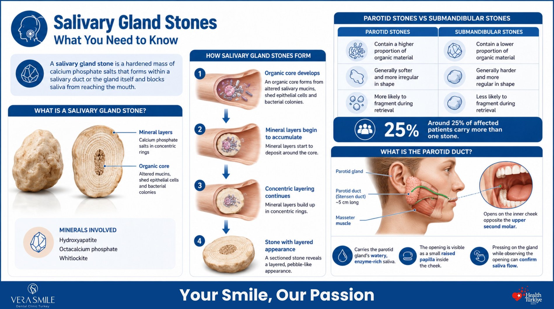
A salivary gland stone is a hardened mass of calcium phosphate salts that forms within a salivary duct or the gland itself and blocks saliva from reaching the mouth. Formation runs in two stages. An organic core develops first, built from altered salivary mucins, shed epithelial cells and bacterial colonies, and mineral layers then accumulate around that core in concentric rings, which gives a sectioned stone the layered appearance of a small pebble (Kraaij et al., 2014). The minerals involved are hydroxyapatite, octacalcium phosphate and whitlockite.
Parotid stones differ from submandibular stones in more than location. They contain a higher proportion of organic material, which makes them softer, more irregular in shape and more likely to fragment during retrieval. Salivary stones belong with tooth decay and gum disease among the dental problems that first present as one-sided facial pain, which is why the diagnosis is often made in a dental chair rather than in a hospital clinic. Around 25% of affected patients carry more than one stone (Kraaij et al., 2014).
What Is the Parotid Duct?
The parotid duct, also called Stensen duct, is a channel roughly 5 cm long that runs forward from the parotid gland across the masseter muscle, pierces the cheek muscle and opens on the inner cheek opposite the upper second molar. It delivers the parotid gland’s contribution to saliva, which is watery and enzyme-rich rather than thick and mucous. That opening is visible in most mouths as a small raised papilla, and pressing on the gland while watching it is the fastest way to confirm whether saliva is flowing on that side.
What Causes Stones to Form in the Parotid Duct?
Stone formation begins when saliva slows down enough for its mineral and protein content to settle out of solution. No single trigger explains every case, and the same patient often carries two or three of the contributing factors at once. Research into stone structure points consistently to a bacterial starting point, since bacteria are recoverable from the core of most analysed stones (Kraaij et al., 2014). The factors that matter clinically are the ones that reduce flow or damage the duct lining.
- Salivary stasis: Any condition that slows the movement of saliva through the duct gives dissolved calcium salts the time they need to precipitate.
- Reduced saliva volume: Chronic dehydration, low fluid intake and illness-related fluid loss concentrate the calcium and phosphate already present in saliva.
- Bacterial biofilm: Colonies growing on the duct wall form the organic core that mineral layers build on, which is why stone cores contain bacterial remnants.
- Duct injury: Cheek biting, orthodontic trauma, a sharp restoration edge or a previous infection leaves scarring that narrows the channel and traps debris.
- Altered salivary proteins: Shifts in mucin composition and pH lower the solubility of calcium phosphate, so the same mineral load precipitates more readily.
- Medication-induced dry mouth: Antihypertensives, anticholinergics, antihistamines, antidepressants and bladder-control drugs all reduce salivary output as a side effect.
These causes compound each other rather than acting alone. A patient on a diuretic who drinks little water during a working day has both reduced volume and increased concentration working against them. That combination explains why stones recur in the same patient after successful removal, and why treatment that ignores the underlying flow problem buys only a few years. Identifying which factors apply to an individual patient is part of the assessment, not an afterthought.
Why Do Fewer Stones Form in the Parotid Than the Submandibular Gland?
The parotid duct is at lower risk because of three anatomical and chemical advantages. Submandibular saliva is more viscous and more alkaline, which favours mineral precipitation, while parotid saliva is thinner and slightly acidic. The submandibular duct also runs uphill from the gland to its opening under the tongue, so saliva travels against gravity, whereas the parotid duct runs a flatter and shorter course. The result is the distribution reported across most case series, with 85% of stones in the submandibular gland and the remainder split between the parotid and, rarely, the sublingual and minor glands (Capaccio et al., 2007).
What Are the Symptoms of a Blocked Parotid Duct?
A blocked parotid duct announces itself at mealtimes. Saliva production surges when eating begins, pressure builds behind the obstruction and the gland swells within minutes. The swelling then subsides as saliva slowly leaks past the stone, producing a cycle that repeats at the next meal. Recognising that pattern is what separates a stone from every other cause of facial swelling.
- Mealtime swelling: The area in front of the ear swells within minutes of eating and settles over the following 1 to 2 hours without treatment.
- Cyclical pain: Discomfort peaks with the swelling and eases between meals, rather than remaining constant through the day.
- Reduced saliva on one side: Pressing the gland produces little or no saliva at the duct opening on the affected cheek.
- A palpable lump: A firm, tender swelling can often be felt along the duct line across the cheek toward the corner of the mouth.
- Foul taste or discharge: Purulent fluid at the duct opening indicates that bacteria have colonised the obstructed duct.
- Restricted jaw opening: Marked swelling limits how far the mouth can open, which is often mistaken for a jaw joint problem.
- Fever and spreading swelling: Systemic signs mean the obstruction has progressed to acute infection of the gland.
Not every stone produces symptoms. A meaningful share are silent and turn up as an unexpected white shadow on a routine panoramic radiograph taken for an unrelated reason. Silent stones still deserve monitoring, because the first symptom many patients experience is an acute infection rather than mild swelling. The moment symptoms shift from a mealtime cycle to constant pain, the situation has changed character.
Warning: Fever, rapidly spreading facial swelling, pus at the duct opening or difficulty opening the mouth points to acute bacterial infection of the gland. This needs same-day medical assessment and antibiotic treatment, not a routine appointment. Delaying care in this situation risks abscess formation and permanent damage to the gland.
Is Mealtime Facial Pain Coming From a Tooth or a Salivary Gland?
The distinguishing question is what provokes the pain. A salivary stone reacts to anything that stimulates saliva, so the pain arrives with the first bite or even at the sight and smell of food. A tooth reacts to temperature, sweetness and biting pressure. A jaw joint reacts to movement, so it hurts on wide opening and chewing rather than on salivation. Getting this wrong leads to root canal treatment on a healthy tooth, which is a common outcome when upper molar pain is assumed to be dental in origin.
Location adds a second layer of information. Parotid pain concentrates in front of and under the ear and often radiates along the jaw, while pain from an upper molar stays closer to the tooth and responds to percussion. A TMJ disorder produces clicking, deviation on opening and tenderness directly over the joint, none of which follow a mealtime swelling cycle.
Clinic Note: Three checks separate these causes at the first examination. The duct opening inside the cheek is inspected while the gland is compressed, to see whether saliva flows on both sides. The duct line is palpated from the gland forward toward the corner of the mouth, since stones sitting near the opening are often felt directly. Suspect teeth are then tested for percussion and thermal response, because a stone leaves those responses normal while an inflamed pulp does not.
How Does Salivary Stone Pain Differ From Toothache and TMJ Pain?
The comparison table sets out the features that separate the three causes at first presentation.
| Feature | Salivary stone | Tooth-origin pain | TMJ disorder |
| Main trigger | Saliva production at mealtimes | Cold, heat, sweetness, biting pressure | Jaw movement and wide opening |
| Timing | Rises within minutes of eating, eases over 1 to 2 hours | Lingers after the trigger is removed | Worse through the day and with use |
| Location | In front of and under the ear, along the cheek | Localised to one tooth, radiating to the jaw | Directly over the joint, in front of the ear |
| Swelling | Visible, cyclical, one-sided | Present only once infection spreads | Absent |
| Saliva flow | Reduced or absent on the affected side | Normal | Normal |
| Response to hydration | Improves with fluids and gland massage | No change | No change |
| First clinician | Dentist for assessment, then ENT or maxillofacial surgery | Dentist | Dentist |
Two features carry most of the diagnostic weight. Cyclical swelling tied to eating points to obstruction almost exclusively, and absent saliva flow on one side confirms it. When both are present, imaging is confirmatory rather than exploratory.
Who Is Most Likely to Develop Salivary Gland Stones?
Salivary stones concentrate in middle age and affect men more often than women. Diagnosis peaks between 30 and 60, and the estimated incidence of symptomatic cases sits at 1 in 10,000 to 1 in 30,000 each year (Escudier & McGurk, 1999). The risk profile is dominated by anything that reduces salivary flow over long periods. Medication is the most common modifiable factor in practice.
- Adults aged 30 to 60: This band accounts for the majority of diagnoses, with cases in children remaining rare.
- Patients on xerostomic medication: Antihypertensives, anticholinergics, antidepressants, antihistamines and bladder-control drugs all suppress salivary output.
- People with low daily fluid intake: Chronic mild dehydration concentrates salivary minerals and slows flow through the duct.
- Patients with Sjögren disease: Autoimmune destruction of glandular tissue reduces flow permanently and raises obstruction risk.
- Patients after head and neck radiotherapy: Radiation damage to glandular tissue reduces saliva volume and alters its composition.
- Smokers: Smoking alters salivary composition and bacterial load in the duct system.
- Patients with gout or kidney disease: Both conditions are reported alongside sialolithiasis, although the mechanism is not established.
Carrying a risk factor does not predict a stone, and most people in these groups never develop one. What the profile does change is how facial swelling should be interpreted. In a patient on three xerostomic medications, one-sided mealtime swelling should be treated as an obstruction until imaging says otherwise. In the same patient, a stone that has already been removed warrants a flow-focused prevention plan rather than a discharge.
How Are Parotid Duct Stones Diagnosed?
Diagnosis moves from the clinical examination to imaging, and stops as soon as the stone is located and measured. Those two pieces of information, size and position, determine every treatment decision that follows. Stones sitting near the duct opening are often identified without any imaging at all. Deeper stones and stones within the gland itself need cross-sectional imaging to plan retrieval.
- Clinical examination: Palpating the duct line and compressing the gland while watching the duct opening identifies distal stones and confirms reduced flow.
- Panoramic radiograph: A dental panoramic film detects denser stones as an unexpected radio-opaque shadow, which is how many silent stones are first found.
- Ultrasound: Ultrasound is the first-line imaging choice, detects stones larger than 2 mm and involves no radiation, which makes it suitable for repeat monitoring (Capaccio et al., 2007).
- Computed tomography: CT resolves small and poorly calcified stones that plain imaging misses, and maps position within the gland before surgery.
- Diagnostic sialendoscopy: A sub-millimetre endoscope passed into the duct shows the stone, the duct wall and any stricture directly, and treatment can follow in the same session.
Density decides what plain imaging can achieve. Roughly 80% of submandibular stones and 60% of parotid stones contain enough mineral to appear on plain radiography, which means a clear film never rules out a stone (Capaccio et al., 2007). Parotid stones are the ones most often missed, because their higher organic content makes them less radio-opaque. A negative radiograph in a patient with cyclical mealtime swelling is a reason to order ultrasound, not a reason to stop looking.
Can a Salivary Stone Be Seen on a Dental X-Ray?
Yes. Dense stones appear on a panoramic radiograph as a well-defined radio-opaque shadow overlying the jaw or the soft tissue of the cheek, and a proportion of stones are discovered this way in patients attending for unrelated dental treatment. Parotid stones are harder to catch than submandibular ones because they carry less mineral. A stone that does not appear on the film is not excluded, so persistent one-sided mealtime swelling should be followed with ultrasound regardless of the radiographic result.
How Are Salivary Gland Stones Treated?
Treatment escalates only as far as the stone requires, and the goal at every step is keeping the gland. That represents a genuine shift in practice. Gland removal was once the standard answer to a recurrent stone, whereas minimally invasive techniques now preserve the gland in the large majority of cases. Stone size and position, rather than symptom severity, decide which step applies.
- Conservative management: Increased fluid intake, sour sweets as a salivary stimulant, gland massage toward the duct opening, warm compresses and anti-inflammatory medication move small stones out without intervention.
- Transoral retrieval and papillotomy: A stone lodged close to the duct opening is retrieved through the mouth under local anaesthesia, with the duct opening widened where access requires it.
- Sialendoscopic retrieval: An endoscope is passed into the duct and the stone is captured with a basket or grasping forceps, which suits parotid stones under about 3 mm and submandibular stones under about 4 mm (Marchal & Dulguerov, 2003).
- Lithotripsy-assisted removal: Laser or extracorporeal shockwave energy fragments a stone too large to pass through the duct, and the fragments are then flushed or retrieved endoscopically.
- Combined endoscopic and transfacial approach: Large or impacted parotid stones are removed through a small external incision guided by the endoscope, achieving stone removal in 99% of cases with gland preservation in effectively all of them (Roland et al., 2017).
- Gland removal: Excision is reserved for a gland already destroyed by repeated infection and scarring, where removing the stone would leave non-functioning tissue behind.
Outcome data supports the conservative-first sequence. Pooled analysis across 9,043 sialendoscopic procedures reports overall success of 80.9%, rising to 89.6% where a stone is the cause and reaching 81.2% in parotid glands (Beumer et al., 2024). For parotid stones treated with sialendoscopy and lithotripsy across 1,285 patients, mean success was 88.7% with no major complications recorded (Galdermans & Gemels, 2020). Those figures make gland removal difficult to justify as a first response to a first stone.
What Is Sialendoscopy?
Sialendoscopy is a minimally invasive procedure that passes an endoscope under 1 mm in diameter into the salivary duct to inspect and treat the duct from inside. The duct opening is dilated, the endoscope is advanced along the channel, and the stone is located, captured and withdrawn in the same session. Duct narrowing can be widened and chronically inflamed ducts irrigated during the same procedure. Diagnostic sessions are performed under local anaesthesia with immediate discharge, while retrieval of deeper stones often requires general anaesthesia.
Which Treatment Suits Which Stone Size and Location?
Stone dimensions and position map onto specific techniques, and the table sets out that relationship.
| Stone size and position | First-line approach | Gland preservation |
| Under 3 mm, near the duct opening | Conservative measures, then transoral retrieval | Very high |
| Under 3 mm parotid, mid-duct | Sialendoscopic retrieval | Very high |
| 3 mm to 7 mm, mid-duct | Sialendoscopy with laser or shockwave fragmentation | High |
| Over 7 mm or impacted, parotid | Combined endoscopic and transfacial approach | High (Roland et al., 2017) |
| Any size, gland already fibrotic | Gland excision | Not achievable |
Size thresholds are guides rather than fixed cut-offs. A soft, irregular parotid stone slightly over the limit may still fragment and pass, while a hard stone within the limit may resist retrieval. Duct diameter at the narrowest point matters as much as the stone itself.
Can a Salivary Gland Stone Be Removed at Home?
No. Probing the duct opening, squeezing the gland forcefully or attempting to dig a stone out with an instrument risks lacerating the duct, and the scar that follows produces a stricture that obstructs saliva permanently even after the stone is gone. What can be done at home is supporting flow so that a small stone passes on its own. Where symptoms recur over more than a week, or where swelling stops resolving between meals, self-care has failed and the stone needs locating.
Tips for Patients: Drink water steadily through the day rather than in large single volumes, since consistent flow matters more than total intake. Suck a sugar-free sour sweet before meals to stimulate saliva and help flush the duct. Massage the gland with firm strokes directed forward toward the duct opening, never backward toward the gland. Apply a warm compress for 10 minutes before massaging, and stop immediately if swelling becomes hot, tense or feverish.
What Happens If a Salivary Stone Is Left Untreated?
An untreated stone rarely stays static. Each obstruction episode leaves the duct inflamed and the gland partly stagnant, and stagnant saliva is a favourable environment for oral bacteria. The sequence that follows is predictable, and it is progressive rather than reversible. Stones account for the majority of obstructive salivary gland disease, which makes this the most common route to losing a salivary gland.
- Recurrent bacterial sialadenitis: Bacteria ascend the obstructed duct and infect the gland, producing pain, pus and fever that returns with each episode.
- Abscess formation: Untreated infection can localise into a collection requiring drainage, which is uncommon but serious.
- Ductal stricture: Repeated inflammation scars the duct wall and narrows the channel, so obstruction continues after the original stone is removed.
- Glandular fibrosis and atrophy: Persistent inflammation replaces functioning secretory tissue with scar tissue, and that loss of function does not recover.
- Gland removal: A gland reduced to fibrous tissue no longer contributes saliva, and excision becomes the only remaining option.
The interval between a first episode and irreversible damage varies from months to years, which is exactly what makes the condition easy to postpone. Patients who manage each episode with fluids and painkillers often present years later with a gland that can no longer be saved. Treating the stone early is a gland-preservation decision, not a comfort decision. That distinction is worth making explicit to any patient inclined to wait it out.
How Can Salivary Gland Stones Be Prevented?
Prevention targets salivary flow, because stones form where saliva stagnates. This matters most for patients who have already had one stone removed, since the conditions that produced the first stone remain in place afterwards. None of these measures guarantees prevention, and each addresses a specific link in the formation chain. Reviewing medication is often the single highest-value step.
- Steady fluid intake: Drinking consistently through the day maintains salivary volume more effectively than large intermittent volumes.
- Medication review: Discussing xerostomic drugs with the prescribing clinician can identify alternatives with less effect on salivary output.
- Active dry mouth management: Saliva substitutes, sugar-free chewing gum and prescribed stimulants restore flow in patients with measured hypofunction.
- Oral hygiene control: Reducing bacterial load in the mouth lowers the bacterial population available to seed a stone core, which is why dental hygiene belongs in a stone-prevention plan.
- Smoking cessation: Stopping smoking improves salivary composition and reduces the duct inflammation associated with tobacco use.
Recurrence is the reason prevention deserves attention rather than a closing paragraph. A patient who has formed one stone has demonstrated the biochemical and anatomical conditions for forming another. Post-treatment follow-up that checks salivary flow and reviews medication catches the second stone while it is still small enough for conservative management. That is a considerably better position than discovering it during an acute infection.
What Does Recovery After Stone Removal Involve?
Recovery from endoscopic or transoral stone removal is short, and most patients return to normal eating within a day. Transient swelling of the gland is expected rather than a complication, since the duct has been dilated and irrigated during the procedure. The recovery plan is built around keeping the duct open while it heals, which means fluids and massage rather than rest. Full soft-tissue healing of the duct takes several weeks.
- First 4 hours: Fluids are resumed shortly after the procedure, and mild discomfort and one-sided swelling are expected during this period.
- First 24 hours: Swelling of the treated gland peaks and settles substantially within the same day, supported by cold compresses and prescribed analgesia.
- First week: Antibiotics are completed where prescribed, soft foods are preferred and gland massage begins as instructed to keep saliva moving through the healing duct.
- Weeks 2 to 4: High fluid intake and daily gland massage continue for roughly 1 month, which is the period when a stricture would form if the duct were left stagnant.
- After the first month: A follow-up review confirms that flow has returned, and imaging is repeated where a second stone or a residual fragment is suspected.
The signs worth watching for are the ones that suggest the duct has narrowed rather than healed. Swelling that returns at mealtimes after an initial recovery, or saliva that remains absent on the treated side, both warrant reassessment rather than waiting. Reported complications of endoscopic treatment are minor and include transient swelling, sialocele and duct stricture. Major complications are not a feature of these techniques in the published series (Roland et al., 2017).
FAQ
Yes. Stones under about 2 mm sitting near the duct opening frequently pass without intervention when saliva flow is stimulated with fluids, sour sweets and gland massage. Patients sometimes notice a small gritty fragment in the mouth as the obstruction clears. Stones that have not moved after a week of consistent self-care are unlikely to pass unaided.
Yes. Bacteria multiplying in stagnant saliva behind an obstruction produce an unpleasant odour, and purulent discharge from the duct opening adds a distinctly foul taste. Reduced saliva volume compounds the problem, since saliva is the mouth’s primary self-cleaning mechanism. Both resolve once the obstruction is cleared and any infection is treated.
No. Salivary stones form through a separate mechanism involving salivary proteins and bacterial nucleation, and having one condition does not predict the others. Kidney disease and gout are reported alongside sialolithiasis in some series, although a causal link is not established (Kraaij et al., 2014). Salivary stones are not treated as a marker for stone disease elsewhere in the body.
Yes. The anatomical and salivary conditions that produced the first stone remain in place once it has been removed, and recurrence in the same gland is well recognised. This is why hydration, medication review and flow monitoring form part of post-treatment care rather than general advice. Recurrent stones caught early remain candidates for conservative management.
No. Viral infection such as mumps, bacterial sialadenitis without a stone, duct stricture, autoimmune conditions including Sjögren disease and salivary gland tumours all cause gland swelling. Stones are the most common cause of obstructive swelling, and the mealtime cycle is what distinguishes them. Swelling that is constant rather than cyclical, or that presents as a firm painless mass, needs a different line of investigation.
Yes. Salivary stones are uncommon before adulthood, and paediatric cases are documented in both the submandibular and parotid ducts. Management follows the same escalation, with a stronger preference for conservative measures and ultrasound rather than radiographic imaging. Recurrent one-sided facial swelling in a child that follows meals should be assessed rather than attributed to a viral cause by default.
A salivary stone forms inside a duct, while tartar forms on the outer surface of a tooth. Both are calcium phosphate deposits and both begin around a bacterial layer, but they sit in different tissues, cause different symptoms and need different treatment. Tartar is scraped away during a routine cleaning. A stone lodged in a duct cannot be reached that way, and neither condition causes the other.
Beumer, C., et al. (2024). Success rate of sialendoscopy, a systematic review and meta-analysis. Oral Diseases, 30(4), 1866–1878.
Capaccio, P., Torretta, S., Ottaviani, F., Sambataro, G., & Pignataro, L. (2007). Modern management of obstructive salivary diseases. Acta Otorhinolaryngologica Italica, 27(4), 161–172.
Escudier, M. P., & McGurk, M. (1999). Symptomatic sialoadenitis and sialolithiasis in the English population, an estimate of the cost of hospital treatment. British Dental Journal, 186(9), 463–466.
Galdermans, M., & Gemels, B. (2020). Success rate and complications of sialendoscopy and sialolithotripsy in patients with parotid sialolithiasis, a systematic review. Journal of Oral and Maxillofacial Surgery, 78(6), 940–948.
Kraaij, S., Karagozoglu, K. H., Forouzanfar, T., Veerman, E. C. I., & Brand, H. S. (2014). Salivary stones, symptoms, aetiology, biochemical composition and treatment. British Dental Journal, 217(11), E23.
Marchal, F., & Dulguerov, P. (2003). Sialolithiasis management, the state of the art. Archives of Otolaryngology–Head & Neck Surgery, 129(9), 951–956.
Roland, L. T., Skillington, S. A., & Ogden, M. A. (2017). Sialendoscopy-assisted transfacial removal of parotid sialoliths, a systematic review and meta-analysis. The Laryngoscope, 127(11), 2510–2516.