
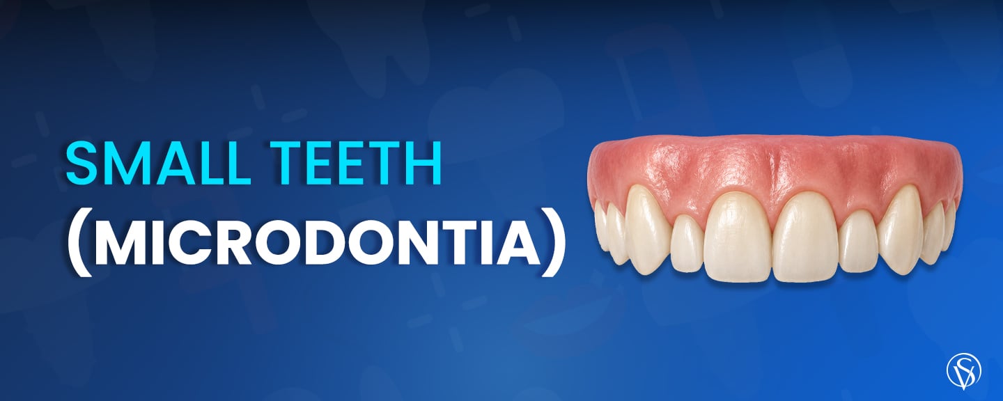
Microdontia is a developmental condition in which one or more teeth are smaller than the normal size range for that tooth type. It is a tooth size anomaly fixed at the moment the tooth forms, not a result of decay, wear, or grinding. Three forms exist. True generalized microdontia affects the whole dentition and is rare, relative generalized microdontia describes normally sized teeth that look small in a large jaw, and localized microdontia affects one or a few teeth.
The classic presentation is a narrow, cone-shaped upper lateral incisor known as a peg lateral. A twin family study of 1,267 people recorded microdontia in 3.00% of participants (Jeong et al., 2015). Microdontia is not a disease and does not threaten tooth survival. The issues it raises are spacing, hygiene access, and proportion. Treatment is elective, and it ranges from composite bonding on a single tooth to veneers, crowns, or orthodontic space redistribution combined with restoration for dental problems.
Key Points
- Microdontia is a developmental anomaly in which one or more teeth form abnormally small for their tooth type, and it describes size rather than disease.
- The localized form accounts for most cases and affects the upper lateral incisor most often, giving the narrow cone shape known as a peg lateral.
- A twin family study of 1,267 people recorded microdontia in 3.00% of participants (Jeong et al., 2015), while the pooled prevalence of peg-shaped upper lateral incisors is 1.8% (Hua et al., 2013).
- Crown size is fixed during tooth formation and is driven mainly by genetics, so microdontia cannot develop later in life and cannot be prevented once the tooth has formed.
- A peg lateral on one side carries a 55% chance of a congenitally missing lateral incisor on the other side (Hua et al., 2013), which is why the opposite side is radiographed before the visible tooth is treated.
- Composite bonding restores an isolated small tooth in one visit with little or no enamel removal, veneers suit several undersized front teeth, and crowns are reserved for teeth that cannot retain a partial-coverage restoration.
What Is Microdontia?
Microdontia is an anomaly of tooth size in which a tooth measures abnormally small relative to the normal range for that tooth type and relative to the patient’s jaw. The term describes size and shape, not health. A microdont tooth has normal enamel, normal dentin, and a normal pulp, and it can be caries-free and fully functional at a reduced size. The condition is diagnosed by clinical measurement and radiographic comparison against the contralateral tooth and against population norms for mesiodistal crown width. Restoring the proportion of small teeth sits within cosmetic dentistry, and the restoration chosen depends on how many teeth are affected and how much width has to be added.
What Do Small Teeth Look Like?
Small teeth present as short or narrow crowns with visible gaps on either side. A peg lateral tapers toward the biting edge and takes a cone or wedge outline instead of the rectangular shape of a normal lateral incisor. In generalized forms the whole arch looks under-scaled against the lips and face, and the teeth may sit with spacing throughout rather than in contact. One distinction matters for treatment planning. Teeth that are genuinely small differ from teeth that only appear small because gum tissue covers part of the crown, a situation seen in altered passive eruption and in a gummy smile. The first case needs the tooth made larger, and the second needs the gum line repositioned, so measuring the crown rather than judging the smile is what separates the two.
What Are the Types of Microdontia?
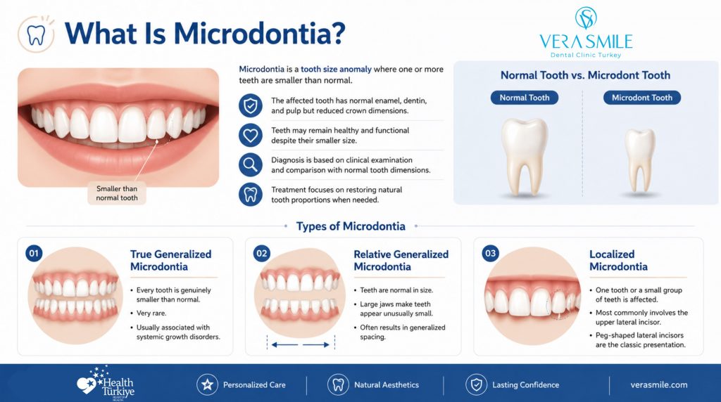
Microdontia is classified by how many teeth are affected and by whether the reduction in size is absolute or apparent. The three recognised forms carry different causes and different treatment implications, and the localized form accounts for the large majority of cases seen in practice. Correct classification changes the plan, because a single peg tooth is a restorative problem while a whole dentition of small teeth is a proportion and space management problem. The three types are:
- True generalized microdontia: Every tooth in the dentition is genuinely smaller than normal, a rare presentation associated with pituitary dwarfism and other disorders of systemic growth.
- Relative generalized microdontia: Teeth of normal size appear small because the jaws and dental arches are larger than average, which produces generalized spacing without any true reduction in crown dimensions.
- Localized microdontia: One tooth or a small group of teeth is affected, most often the maxillary lateral incisor and the upper third molar, and the affected lateral incisor commonly takes the peg form.
Localized microdontia of the upper lateral incisor is well quantified. A meta-analysis of 36 studies found a pooled prevalence of peg-shaped maxillary permanent lateral incisors of 1.8%, rising to 2.7% among orthodontic patients, with women affected 1.35 times more often than men (Hua et al., 2013).
What Causes Microdontia?
Microdontia is caused by disruption of tooth size determination during the early stages of tooth formation, and genetic factors carry the greater share of that disruption. Crown dimensions are set during the bell stage of odontogenesis, years before the tooth erupts, which is why the condition cannot develop later in life and cannot be prevented once the tooth has formed. Both inherited and acquired influences have been documented, and in many patients more than one is present. The recognised causes are:
- Genetic and hereditary factors: Family clustering is strong, and a twin family study estimated an additive genetic effect of 0.38 for hypodontia and microdontia pooled, confirming inherited transmission of tooth size anomalies (Jeong et al., 2015).
- Developmental disruption during odontogenesis: Interference with the signalling that sets crown size produces a smaller tooth germ, and the effect is confined to the teeth forming at that moment.
- Systemic and endocrine conditions: Growth hormone deficiency is associated with altered tooth cell differentiation, and children with isolated growth hormone deficiency showed dental anomalies of number or size in 33% of cases against 4% of healthy controls (Torlińska-Walkowiak et al., 2023).
- Childhood cancer treatment: Chemotherapy and head or neck radiation delivered while permanent teeth are still forming can reduce crown and root dimensions in the teeth developing at the time of treatment.
- Association with missing teeth: Microdontia and hypodontia share a genetic pathway and appear together often, and 55% of people with a peg lateral on one side have a congenitally missing lateral incisor on the other side (Hua et al., 2013).
That last association is the most clinically useful of the group. A peg lateral on one side is a reason to radiograph the opposite side rather than to treat the visible tooth in isolation, because the treatment plan changes entirely when a tooth is absent as well as undersized.
Which Genetic and Syndromic Conditions Are Linked to Microdontia?
Microdontia appears as a feature of several recognised syndromes, and in those cases it presents alongside other dental and systemic findings rather than on its own. Identifying a syndromic pattern matters because it predicts additional anomalies of tooth number, shape, and eruption that affect the sequencing of treatment. The conditions most often reported in association with microdontia are the following.
- Down syndrome: Reduced crown dimensions occur across the dentition together with delayed eruption, hypodontia, and a high frequency of peg-shaped upper lateral incisors.
- Ectodermal dysplasia: Small and conical teeth are a defining oral feature, presenting with severe hypodontia or oligodontia and with reduced alveolar bone development.
- Turner syndrome: Crown size is reduced in both dimensions, with thinner enamel reported across the permanent dentition.
- Cleft lip and palate: Teeth adjacent to the cleft show reduced size and altered shape, and the upper lateral incisor on the cleft side is affected most often.
- Growth hormone deficiency: Anomalies of tooth size and number were recorded in one third of affected children, at a rate significantly above healthy controls (Torlińska-Walkowiak et al., 2023).
A syndromic diagnosis does not rule out restorative treatment, and it does shift the emphasis toward multidisciplinary planning, because tooth number and bone volume have to be assessed before any tooth is prepared.
Clinic Note: A single peg-shaped lateral incisor in an otherwise normal dentition points to isolated localized microdontia. Small teeth across the whole arch, particularly with generalized spacing or delayed eruption, warrant screening for an underlying systemic or syndromic cause before restorative treatment starts.
What Problems Can Microdontia Cause?
Microdontia’s problems are functional and aesthetic rather than pathological, and they follow from the space undersized teeth leave rather than from disease risk. The affected tooth is not weakened, and its lifespan is not shortened. What brings patients to a dental clinic is the gap left on either side of an undersized crown and the proportion of the front teeth, and both are worth treating on their own terms. The effects reported most often are the following.
- Spacing between teeth: Undersized crowns leave gaps that the neighbouring teeth cannot close on their own, and the resulting midline gap is a form of diastema driven by tooth width rather than by a frenal attachment or a missing tooth.
- Food trapping and hygiene demand: Wider interdental spaces hold food and plaque, which raises the daily cleaning burden without raising decay risk on its own.
- Bite and alignment effects: Where several teeth are undersized, arch length exceeds the space the teeth occupy, and adjacent teeth can drift or rotate into the surplus.
- Proportion of the front teeth: Short or narrow crowns break the width-to-height ratio of the upper incisors, which is the reason patients ask about treatment most often.
None of these effects calls for urgent intervention. They set the reason for treatment, and they also set its timing, since a patient content with the appearance of small teeth and able to clean around them needs monitoring rather than restoration.
How Is Microdontia Treated?
Microdontia is treated by adding the missing tooth structure back with a bonded restoration, and the method is chosen by how many teeth are involved, how much width has to be added, and how much sound enamel can be preserved. Treatment is elective in every case. The guiding principle is that the least invasive option capable of producing the required width and shade should be used first, because enamel removed for a restoration cannot be replaced. Where orthodontic space exists in the wrong place, moving teeth before restoring them produces a better result than widening a tooth into a gap it cannot fill. The available options are:
- Composite bonding: Resin composite is added directly to the tooth and shaped in a single appointment, with little or no enamel removal, which makes it the first option for an isolated peg lateral.
- Dental veneers: Thin ceramic facings restore width, length, and shade across several anterior teeth at once, and the approach suits generalized or multi-tooth microdontia where shade change is also wanted.
- Dental crowns: Full-coverage restorations rebuild teeth that are very small or structurally compromised, and they are reserved for cases where a partial-coverage restoration cannot achieve retention.
- Orthodontic treatment before restoration: Aligners or fixed appliances redistribute the spacing so that each small tooth receives an even amount of added width, and this step precedes the restorative phase where several teeth are affected.
- Implant planning where a tooth is also missing: When microdontia coexists with a congenitally absent tooth, the plan combines space management, an implant or a bonded bridge for the missing unit, and a restoration for the undersized one.
Each of these options adds material to the tooth rather than removing it, and the difference between them lies in how much enamel has to be sacrificed to make room for that material.
Which Treatment Is Best for Small Teeth?
The best treatment for small teeth is the one that adds the required width with the least enamel loss, which places composite bonding first for one or two teeth and dental veneers first for multiple anterior teeth. Published survival data supports both as durable choices, and it also shows the trade-off clearly, since ceramic outlasts composite while requiring more tooth preparation.
| Aspect | Composite bonding | Dental veneers | Dental crowns |
| Enamel preparation | None to minimal | Thin facial reduction | Full-coverage reduction |
| Reversibility | Reversible | Largely irreversible | Irreversible |
| Reported survival | 88% pooled and 91% for the direct technique over a 24 to 97 month follow-up (Lim et al., 2023) | 95.5% cumulative survival at 10 years across 6,500 restorations (Alenezi et al., 2021) | 94.7% metal-ceramic and 96.6% lithium disilicate at 5 years (Sailer et al., 2015) |
| Appointments | Single visit | Two or more visits | Two or more visits |
| Repair after damage | Repairable at the chair | Replacement on failure | Replacement on failure |
| Best suited to | One or two small teeth, isolated peg lateral | Several small anterior teeth needing width and shade change | Very small or structurally weak teeth |
The figures place ceramic ahead of composite on longevity by a clear margin, and they do not settle the choice on their own. A reversible restoration on an intact tooth in a patient in their twenties preserves options that a veneer preparation closes off permanently, which is why sequencing from the least invasive option upward is the approach applied at Vera Smile, with dental crowns reserved for teeth that cannot retain a partial-coverage restoration.
Can Microdontia Be Treated in Children?
No. Definitive restoration of small teeth waits until the permanent dentition is complete and jaw growth has finished, because a restoration built onto a growing arch will not match the final tooth position, gum level, or arch width. What is appropriate during childhood is assessment, monitoring, and space management, which protect the options available once growth is complete. Interim measures are conservative and reversible. Provisional composite build-ups can be added and adjusted as the child grows, and orthodontic space maintenance keeps the room a definitive restoration will need. Assessment during the mixed dentition years falls within pediatric dentistry and produces the radiographic record that later planning depends on.
When Should a Child Be Referred for a Microdontia Assessment?
Referral for assessment is warranted when any of the following applies.
- A peg-shaped or undersized tooth is present, since the contralateral side needs radiographic checking for a missing tooth.
- Several teeth are small, which raises the question of an underlying systemic or syndromic cause.
- Spacing is increasing or adjacent teeth are drifting into the surplus space.
- A permanent tooth has failed to appear within the expected window for its position.
Small teeth in a child should be assessed by a dentist rather than treated on the basis of appearance. Irreversible preparation of a tooth before growth is complete can compromise the definitive result, and outcomes in a growing patient cannot be predicted with the certainty possible in an adult.
How Much Does It Cost to Fix Small Teeth?
Treating small teeth is priced per tooth rather than per case, so the total depends on how many undersized teeth are restored and which material is chosen. Composite bonding sits at the lower end because it is completed in one appointment with no laboratory stage, while ceramic veneers and crowns carry laboratory and material costs on top of clinical time. Istanbul pricing sits at the lower end of the international range for the same ceramic systems and laboratory standards. The per-tooth range for each option appears in the table.
| Treatment | What the fee covers | Range per tooth | Service life |
|---|---|---|---|
| Composite bonding | Consultation, shade selection, direct build-up and finishing in a single visit | €100 to €400 | 3 to 7 years |
| Composite veneer | Chairside or laboratory-made resin facing across the front surface | €100 to €700 | 5 to 7 years |
| Ceramic veneer, porcelain or lithium disilicate | Digital planning, preparation, laboratory fabrication, trial fit and bonding | €250 to €1,500 | 10 to 15 years |
| Crown, zirconia or lithium disilicate or PFM | Preparation, temporary restoration, laboratory fabrication and cementation | €250 to €900 | 10 to 20 years |
Per-tooth pricing means a single peg lateral and six undersized front teeth produce very different totals, and the assessment appointment converts the range into a fixed figure for the individual case. Where an orthodontic phase is needed to redistribute spacing before restoration, that phase is quoted separately from the restorative work. Dental veneers cost varies by ceramic system, with lithium disilicate priced above conventional porcelain for the same tooth. Dental crowns cost spans a wider band, because material options run from porcelain-fused-to-metal at the lower end to gold alloy at the upper end.
What Affects the Cost of Treating Small Teeth?
Four factors move the price within each treatment type, and the number of teeth is the largest of them. Knowing which factors apply makes a quotation readable rather than arbitrary. The factors that change the figure are the following.
- Number of teeth restored: Pricing is per tooth, so the total scales directly with how many undersized teeth are treated, and multi-tooth cases distribute planning and setup fees across several restorations.
- Material selected: Zirconia, lithium disilicate and porcelain-fused-to-metal carry different laboratory costs, and the choice follows the position of the tooth and the shade change required.
- Need for an orthodontic phase: Redistributing spacing before restoration adds a separate treatment phase with its own quotation.
- Presence of a missing tooth alongside the small one: A congenitally absent lateral incisor requires an implant or a bonded bridge in addition to the restoration, which changes both the plan and the total.
Each of these is identified at the assessment appointment, which is why a figure quoted before examination remains an estimate rather than a quotation.
FAQ
Small teeth rarely affect chewing, because the molars carry the chewing load and localized microdontia affects the front teeth and third molars most often. Speech can be affected when spacing between the upper front teeth is wide enough to alter airflow on sibilant sounds. In generalized microdontia, where many teeth are undersized and contacts are absent throughout the arch, chewing efficiency can drop and warrants full assessment.
Yes. Direct composite bonding restores an isolated peg lateral in one appointment with no preparation of the adjacent teeth. Shade matching is the limiting factor, because a composite added to a single tooth has to blend with two untouched neighbours. Where the shade of the whole front segment is also to be changed, treating several teeth together produces a more even result.
Microdontia does not increase decay or gum disease risk directly, and the spacing it creates changes what effective cleaning requires. Wider interdental spaces trap food more readily, and they are also more accessible to interdental brushes than tight contacts are. Risk rises after treatment if restoration margins are not maintained, which makes the margin between an added restoration and the tooth the area to clean carefully.
Small teeth cannot be made biologically larger, and their visible size can be permanently increased with a bonded restoration. Crown dimensions are set during tooth formation and cannot grow afterwards, so all treatment works by adding material to the outside of the tooth. Composite, ceramic veneers, and crowns each restore the missing width, and each requires eventual replacement or repair rather than lasting for life.
Alenezi, A., Alsweed, M., Alsidrani, S., & Chrcanovic, B. R. (2021). Long-term survival and complication rates of porcelain laminate veneers in clinical studies: A systematic review. Journal of Clinical Medicine, 10(5), 1074.
Hua, F., He, H., Ngan, P., & Bouzid, W. (2013). Prevalence of peg-shaped maxillary permanent lateral incisors: A meta-analysis. American Journal of Orthodontics and Dentofacial Orthopedics, 144(1), 97–109.
Jeong, K. H., Kim, D., Song, Y.-M., Sung, J., & Kim, Y. H. (2015). Epidemiology and genetics of hypodontia and microdontia: A study of twin families. The Angle Orthodontist, 85(6), 980–985.
Lim, T. W., Tan, S. K., Li, K. Y., & Burrow, M. F. (2023). Survival and complication rates of resin composite laminate veneers: A systematic review and meta-analysis. Journal of Evidence-Based Dental Practice, 23(4), 101911.
Sailer, I., Makarov, N. A., Thoma, D. S., Zwahlen, M., & Pjetursson, B. E. (2015). All-ceramic or metal-ceramic tooth-supported fixed dental prostheses (FDPs)? A systematic review of the survival and complication rates. Part I: Single crowns (SCs). Dental Materials, 31(6), 603–623.
Torlińska-Walkowiak, N., Majewska, K. A., Sowińska, A., Kędzia, A., & Opydo-Szymaczek, J. (2023). Developmental enamel defects and dental anomalies of number and size in children with growth hormone deficiency. Scientific Reports, 13(1), 14707.